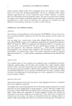
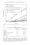
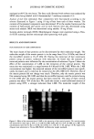
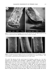
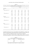
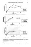
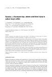
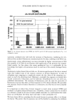
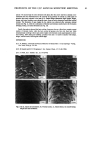
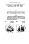
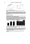
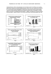
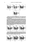
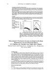
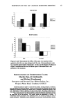
PREPRINTS OF THE 1997 ANNUAL SCIENTIFIC MEETING 55 It is concluded that the perception of skin type is due more to variation in surface oil than a variation in moisture level. In addition, the lack of correlation between moisture and sebum levels suggest that sebum may not be serving as a defense against skin dehydration and a high level of sebum does not influence the effect of a light moisturizer. These results also suggest that individual perception of skin type may not always result in correct product usage. 1. Laufer A. & Dikstein, $., Cosmetics & Toiletries, 111, 91-98, 1996 MODULATION OF INFLAMMATORY REACTION IN THE SKIN: A NEW APPROACH IN THE TREATMENT OF PREMATURE AGING T. Mammone, K. Marenus, E. Pelle, D. Maes Esteb Lauder Laboratories, Melville, NY 11747 Abstract: The human skin is permanently challenged by prooxidative events generated exogeneously by the environment (UV light, smoke), and endogenously by the reaction of the skin's own immune reaction: inflammatory reactions. A significant amount of research has been done over the past years, showing the interest of an anti-oxidant therapy to control the pace of the premature aging process. However, very little work has been done so far to evaluate the activity a topical treatment containing anti-inflammatory compounds in protecting the skin from the damages resulting from the over reaction of the immune system. We propose to review the role played by the inflammatory reactions in the development of the premature aging process, and then to present the available technologies which allows to reduce significantly the progress of inflammatory reactions in the skin. Introduction: The human skin is constantly exposed to environmental insults that challenge it's integrity and function. These insults are in themselves damaging to the skin structure but they also precipitate the immensely complex inflammatory reaction that also if in excess will degrade and atrophy the tissue. Contributing to this wasting induced by the insult is the poor repair function observed in chronically aged skin that limits the replacement of extracellular matrix and cells. We theorize that the struggle between anabolic and catabolic forces will shift with increasing age and result in a more catabolic nature. Therefore the tissue will degrade both from external and internal drives. The cosmetic appearance of aging skin is due to these forces. Methods: Antioxidant ability was measured using a liposome oxidation method. Liposomes composed of phosphatidylcholine are irradiated with UVB or UVA light from Philips FS40 bulbs. In vitro Assays: Arachidonic acid release was monitored by prelabeling for 24 hours followed washing free unincorporated label. After UV irradiation samples of media are counted in scintillation counter. Interleukin-lalpha and TNF-alpha release are measured using commercial available ELISA kits on supernatants from culture media. Collagenase activity was measured using N-2,4 dinitrophenyl-pro-leu- gly-leu-trp-ala-D-ard-amide. This substrate is converted to a fluorescent product which is measured at 360 nm after excitation with 280 nm (1). Elastase activity was measured using the fluorescent substrate N- methyoxysuccinyl-ala-ala-pro-val-amido-4-methyl coumarin (2). Human leukocyte elastase (Sigma Co.) was used in reaction. Enzyme reaction was monitor using fluorescent excitation at 370 nm and emission at 470 nm. Results: UV irradiation of both human skin and artificial skin grown in the lab demonstrated increased levels of lipid peroxidized. Figure 1 shows that 100 mJ/cm2 of UVB increased the amounts of TBA reactive material (lipid peroxides) over time in human skin models. This increased lipid peroxidation was o reduced if skin models were pre- and post- UVB treated with antioxidant mixture of 1 'A vitamin E and 1% vitamin C.

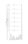
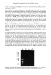
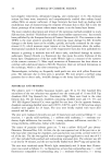
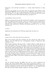
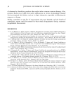
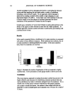
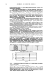
56 JOURNAL OF COSMETIC SCIENCE Figure # 1 Figure # 2 0.4! O.2 0 0 Effect ofAntioxidar• m UVB-In:tmed Peroxidatim ffi UV-B Induced IL - 1 a Release From Skin Model uv-a Human skin has been observed to release interleukin-1 alpha after being irradiated with UV light (3). We have observed similar release of interleukin-I alpha from skin models after UVB irradiation (figure # 2). 250 mJ/cm, 500 mJ/cm2 and 1000 mJ/cm2 increased the media levels of interleukin-! alpha from 300 pg/ml to 500 pg/m1475 pg/ml and 675 pg/ml, respectively. Release of infi•ory Cytokit• UV8 • release ofmNF oc 150 -- 0 50 1011 150 •11 Evaluation of the UV-Induced Activation of Proteases UVB Indur. ad elasia• ar•vity UVB indur. ad colla•nase In living s•xJn mndd a•v•y In IMng stdn 0.7 0 • • 1• 0 • • •b {mgc• uvb (mJ/c• Figure # 3 Figure # 4 Tumor necrosis factor alpha (TNF alpha) has also been shown to be released from human skin (4). We observed release from skin models in culture after treatment with UVB doses of 0 - 200 mJ/cm2 (figure # 3). Both interleukin-1 alpha and TNF alpha have catabolic functions in that they cause the degradation of various tissues and extracellular matrix (5). We observed that UVB increase the enzymatic activity for collagenase and elastase in extracts from human skin models (Figure/t4). UVB doses of 250 mJ/cm2, 500 mJ/cm2 and 1000 mJ/cm2 in collagenase activity between 1.5 - 2 fold over unirradiated controls. Elastase activity was increased by 2 fold at the highest doses of UVB. Conclusion: The inflammatory response of human skin to various environmental insults, such as UV light or cigarette smoke, results in the release of messengers with detrimental effects. Interleukin-I and TNF alpha are well know to increase the catabolic metabolism of a tissue. These inflammatory cytokines will contribute to the degradation and atrophy characteristic ofphotoaged skin. In addition, UV and enviroranental pollutants activity the catalytic enzymes ,collagenase and elastase, that will directly cause atrophy and tissue degradation of exposed skin. Therefore a successful cosmetic intervention for photoaging or premature aging must address this catabolic pathway at multilevels to suppress the detrimental effects. The treatment cosmetic design should incorporate the benefits of antioxidants, protease inhibitors, and inflammatory cytokine release inhibitors. Reference: 1. S.Netze!-Arnett, S.K. Mallya, H.Nagase, H.Birkedal-Hansen, H.E.Van Wart.. Anal. Biochem. 195:86-92 (1991). 2. Castillo, M.J., Nakajima, K., Zimmerman, N., Powers, J.C. Anal. Biochem. 99:53-64 (1979) 3. Kupper, T., Chua, A.O., Flood, P., McGuire, J., Gubler, U.. J.Clin. Invest. 80:430-436 (1987) 4. Kock, A., Schwarz, T., Kirnbauer, R., Urbanski, A., Perry, P., Ansel, J.C., Luger, T. (1990) J. Experimental Med. 172: ! 609- ! 6 ! 4 (! 990) 5. Postethwaite, A.E., Lachman, L.B., Mainardi, C., Kang, A.H.,. J.Exp. Med. 157:801-806 (1983)
Purchased for the exclusive use of nofirst nolast (unknown)
From: SCC Media Library & Resource Center (library.scconline.org)