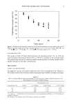
1999 ANNUAL SCIENTIFIC MEETING 71 A CELL-BASED PIGMENTATION ASSAY SUITABLE FOR SCREENING OF COSMETIC RAW MATERIALS Brian Costello, Ph.D. Collaborative Laboratories, Stony Brook, NY 11790 There is substantial interest in evaluating cosmetic raw materials for effects on melanin production. Since tyrosinase activity is considered rate limiting in melanogenesis, potential skin lightening materials are often screened by measuring their effects on this enzyme. However, melanogenesis involves a series of additional reactions, some of which may be amenable to modulation. In addition, the ability of an inhibitor to be effective in actual use can be limited by its skin and cell penetration properties. While panel testing is the only definitive method to determine efficacy under use conditions, large scale screening of potential actives in vivo is impractical and expensive. Therefore, a system was needed that would allow preliminary screening of actives and formulations prior to panel testing of only the most promising materials. To address this problem, we've experimented with a tiered in vitro screening system that incorporates a cell-based whitening/tanning assay as an intermediate between initial tyrosinase inhibition screening and an in vitro test using epidermal equivalent tissues that contain active melanocytes (Melanoderm). Stage 1. The first test phase is tyrosinase activity screening using either tyrosine or DOPA as substrate and measuring dopachrome generation spectrophotometrically. This method is quick and extremely cost- effective but is limited in that it only detects effects on the initial steps of melanogenesis. Nevertheless, this procedure has been used widely and successfully to identify new raw materials as potential skin lightening agents. Stage 2. The second stage of the screening regimen utilizes cultured melanocytes in a procedure similar to that outlined by Dooley et al (1). Cloudman S91 melanoma cells are cultured in multiwell plates in the presence of melanocyte stimulating hormone (ct-MSH) to promote melanogenesis. The cells are simultaneously exposed to varying concentrations of test material. (A preliminary cytotoxicity measurement may be performed and the results of this test, in conjunction with Stage 1 tyrosinase inhibition data, facilitate determination of the initial concentration range to test.) After a 6-day treatment period, the melanin content of the cultures is determined by detergent lysis of the cells followed by peroxide solubilization of melanin and optical density measurement. Cytotoxicity is assessed using similarly treated cultures of melanocytes and/or another skin cell type. By challenging keratinocytes or fibroblasts in addition to melanocytes, melanocyte-specific toxicity can be detected. Viability is measured by dye binding methods using either neutral red or crystal violet. Since this Stage 2 test system will demonstrate activity only for materials that penetrate the cell membrane, it's also useful for rapid screening of delivery vehicles. Although not as simple as the tyrosinase activity screen, the cell-based test is still amenable to rapid, large-scale exploratory screening. Stage 3. The f'mal screening phase uses "Melanoderm" epidermal equivalent tissues developed by MatTek Corporation. These tissues comprise a stratified epidermis containing active melanocytes in the basal layer and a stratum corneum at the air interface Because of its stratum corneum, Melanoderm also is useful for testing finished formulations. Materials are tested by applying an aliquot to the surface every other day for a total treatment period of 7-10 days. The culture medium contains both ct-MSH and fibroblast growth factor and very pronounced darkening of control tissues is observed. Tissue melanin content is scored by enzymatic digestion, followed by peroxide solubilization and spectrophotometric measurement. Simultaneous toxicity measurements can be performed on tissues treated in parallel using the MTT method. We have screened several known or potential melanogenesis inhibitors through this system in order to determine the predictive value of each test stage. Most of the test materials we used had been initially characterized as tyrosinase inhibitors, and in some cases performance in the cell and tissue stages of screening was dependent on effective delivery. A particular focus of this study was to determine whether the cell-based test was a sufficient f'mal stage for in vitro screening of raw materials. 1. T.P. Dooley, R.C. Gadwood, K. Kilgore & L.M. Thomasco, Skin Pharmacology, 7, ! 88-200 (1994)
Purchased for the exclusive use of nofirst nolast (unknown)
From: SCC Media Library & Resource Center (library.scconline.org)