J. Cosmet. Sci., 58, 11-17 Oanuary/February 2007)
Structural analysis of the cell membrane complex in the
human hair cuticle using microbeam X-ray diffraction:
Relationship with the effects of hair dyeing
TAKAFUMI INOUE, YOSHIMICHI IWAMOTO,
NOBORU OHTA, KATUAKI INOUE, and NAOTO YAGI,
Basic Research Laboratory, Kanebo Cosmetics, Inc., 5-3-28 Kotobuki-cho,
Odawara, 250-0002, Japan (T.I.), Beauty Care Laboratory, Kanebo
Home Products Ltd., 134 Goudo-cho, Hododaya-ku, Yokohama,
240-0005, Japan (Y.I.), Japan Synchrotron Radiation Research
Institute UASRI/SPring-8)1 Hyogo, 679-51981 Japan (N.0.1 K.I.,
N.Y.)
Accepted for publication September 20, 2006.
Synopsis
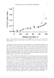
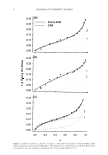
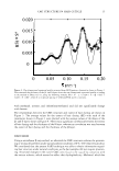
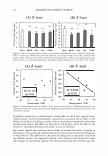
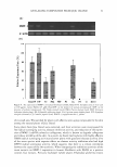
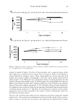
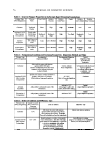
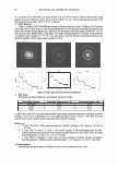
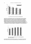
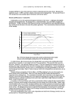
This article deals with the structure of the cell membrane complex (CMC) in the human hair cuticle. The
microbeam X-ray provided a pattern of small-angle scattering from the CMC in the cuticle with no sample
preparations, including slicing and pre-staining of hair. The thickness of the 13- and 8-layers, substructure
in CMC, was estimated by analysis of the scattering pattern. We used hair samples extracted with several
solvents, and found that solvent extraction changed the thickness of the 13- and 8-layers in a manner
dependent on the type of solvent. Extraction of hair with solvent was also shown to have effects on the extent
of dyeing. There was a high correlation between the extent of dyeing and the thickness of the 8-layer, i.e.,
a thin layer tended to show a high amount of dyeing, whereas there was no significant correlation between
the thickness of the 13-layer and the extent of dyeing.
INTRODUCTION
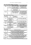
The cuticle is the outermost layer of a hair fiber and is made up of a stack of approxi-
mately ten sheet-like cells that is 0.5 µm thick and roughly 60 µm square. The surface
of each cell is covered by a thin layer of lipids called the 13-layer (2.5 to 4.0 mm thick),
and these lipids are separated between adjacent cells by the 8-layer (15 to 18 nm thick),
which acts as an intercellular cement (1). The exact composition of the 8-layer is still
unknown however, it has been proposed to contain non-keratinous proteins (2). This
lipid-protein cement-lipid structure is called the cell membrane complex (CMC) and is
the only substructure that continuously fills the intercellular spaces of hair fibers.
Address all correspondence to Takafumi Inoue.
11
Structural analysis of the cell membrane complex in the
human hair cuticle using microbeam X-ray diffraction:
Relationship with the effects of hair dyeing
TAKAFUMI INOUE, YOSHIMICHI IWAMOTO,
NOBORU OHTA, KATUAKI INOUE, and NAOTO YAGI,
Basic Research Laboratory, Kanebo Cosmetics, Inc., 5-3-28 Kotobuki-cho,
Odawara, 250-0002, Japan (T.I.), Beauty Care Laboratory, Kanebo
Home Products Ltd., 134 Goudo-cho, Hododaya-ku, Yokohama,
240-0005, Japan (Y.I.), Japan Synchrotron Radiation Research
Institute UASRI/SPring-8)1 Hyogo, 679-51981 Japan (N.0.1 K.I.,
N.Y.)
Accepted for publication September 20, 2006.
Synopsis
This article deals with the structure of the cell membrane complex (CMC) in the human hair cuticle. The
microbeam X-ray provided a pattern of small-angle scattering from the CMC in the cuticle with no sample
preparations, including slicing and pre-staining of hair. The thickness of the 13- and 8-layers, substructure
in CMC, was estimated by analysis of the scattering pattern. We used hair samples extracted with several
solvents, and found that solvent extraction changed the thickness of the 13- and 8-layers in a manner
dependent on the type of solvent. Extraction of hair with solvent was also shown to have effects on the extent
of dyeing. There was a high correlation between the extent of dyeing and the thickness of the 8-layer, i.e.,
a thin layer tended to show a high amount of dyeing, whereas there was no significant correlation between
the thickness of the 13-layer and the extent of dyeing.
INTRODUCTION
The cuticle is the outermost layer of a hair fiber and is made up of a stack of approxi-
mately ten sheet-like cells that is 0.5 µm thick and roughly 60 µm square. The surface
of each cell is covered by a thin layer of lipids called the 13-layer (2.5 to 4.0 mm thick),
and these lipids are separated between adjacent cells by the 8-layer (15 to 18 nm thick),
which acts as an intercellular cement (1). The exact composition of the 8-layer is still
unknown however, it has been proposed to contain non-keratinous proteins (2). This
lipid-protein cement-lipid structure is called the cell membrane complex (CMC) and is
the only substructure that continuously fills the intercellular spaces of hair fibers.
Address all correspondence to Takafumi Inoue.
11