CMC STRUCTURE IN HAIR CUTICLE 13
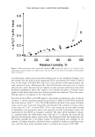
atmosphere of 30 ° C and 50% relative humidity. A high-flux beam emitted from a
helical undulator (A. =0.083 nm) was focused with two mirrors laid horizontally and
vertically (8). In an experimental hutch, an X-ray beam 5 µm in diameter was produced
behind two pinholes, the first 5 µm in diameter and the second 100 µm in diameter. The
sample-to-detector distance was set at approximately 2.3 m. The reciprocal spacing (S)
was calibrated by a spacing of 4.894 nm for lead stearate. The X-ray diffraction profile
was recorded by a two-dimensional detector with an exposure time of 1 second, using an
X-ray image intensifier coupled to a cooled CCD camera (1024 x 1024 pixels). The
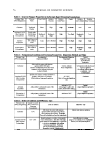
thickness of the �-layer and 3-layer was estimated using the method of Ohta et al. (7).
RESULTS
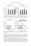
EFFECTS OF SOL VENT EXTRACTION ON HAIR DYEING
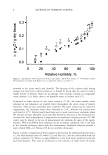
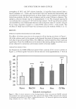
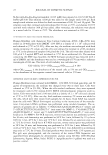
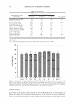
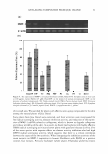
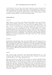
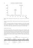
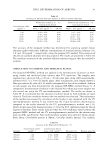
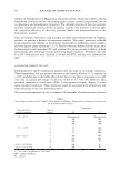
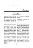
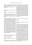
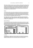
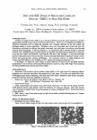
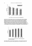
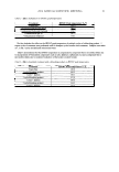
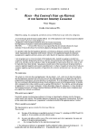
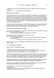
The effects of solvent extraction on the properties of hair dyeing are shown in Figure 1.
All of the solvents used in the present study elevated the extent of dyeing as compared
with that of non-extracted hair fibers. The differences seemed slight but have statistical
significance. There were differences in the extent of hair dyeing among the solvents used,
as hair subjected to extraction with hexane tended to become smaller.
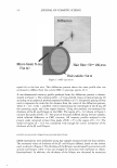
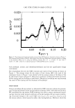
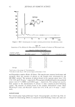
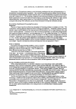
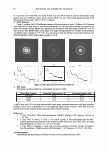
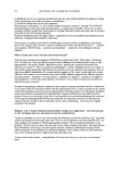
DIFFRACTION FROM CUTICLE
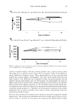
An illustration of a SAXS diffraction pattern from a portion of the cuticle is shown in
Figure 2, in which signals vertical to the hair axis are tilted approximately 3° with an
46 ***
45
44
***
]..__,,,
+'43
C
Q.) .µ 42 X
Q)
b.O 41 -�
Q) 40 �
0
39
38
None MeOH Ace Hex Cl/Me
Solvent extraction
Figure 1. Effect of solvent extraction on hair dyeing properties. None: non-extracted. MeOH: extracted
with methanol. Ace: extracted with acetone. Hex: extracted with hexane. Cl/Me: extracted with a mixture
of chloroform and methanol (2:1). Mean ± standard derivation (n =8). :-itat1st1cal significance was analyzed
using a Dunnett test. *p 0.05, ***p 0.001.
atmosphere of 30 ° C and 50% relative humidity. A high-flux beam emitted from a
helical undulator (A. =0.083 nm) was focused with two mirrors laid horizontally and
vertically (8). In an experimental hutch, an X-ray beam 5 µm in diameter was produced
behind two pinholes, the first 5 µm in diameter and the second 100 µm in diameter. The
sample-to-detector distance was set at approximately 2.3 m. The reciprocal spacing (S)
was calibrated by a spacing of 4.894 nm for lead stearate. The X-ray diffraction profile
was recorded by a two-dimensional detector with an exposure time of 1 second, using an
X-ray image intensifier coupled to a cooled CCD camera (1024 x 1024 pixels). The
thickness of the �-layer and 3-layer was estimated using the method of Ohta et al. (7).
RESULTS
EFFECTS OF SOL VENT EXTRACTION ON HAIR DYEING
The effects of solvent extraction on the properties of hair dyeing are shown in Figure 1.
All of the solvents used in the present study elevated the extent of dyeing as compared
with that of non-extracted hair fibers. The differences seemed slight but have statistical
significance. There were differences in the extent of hair dyeing among the solvents used,
as hair subjected to extraction with hexane tended to become smaller.
DIFFRACTION FROM CUTICLE
An illustration of a SAXS diffraction pattern from a portion of the cuticle is shown in
Figure 2, in which signals vertical to the hair axis are tilted approximately 3° with an
46 ***
45
44
***
]..__,,,
+'43
C
Q.) .µ 42 X
Q)
b.O 41 -�
Q) 40 �
0
39
38
None MeOH Ace Hex Cl/Me
Solvent extraction
Figure 1. Effect of solvent extraction on hair dyeing properties. None: non-extracted. MeOH: extracted
with methanol. Ace: extracted with acetone. Hex: extracted with hexane. Cl/Me: extracted with a mixture
of chloroform and methanol (2:1). Mean ± standard derivation (n =8). :-itat1st1cal significance was analyzed
using a Dunnett test. *p 0.05, ***p 0.001.