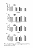
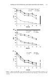
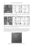
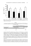
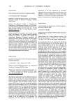
EFFECTS OF PLACENTAL EXTRACT ON FIBROBLAST PROLIFERATION 197 and an electron-coupling reagent was added to each well and catalyzed by mitochondrial dehydrogenase enzymes in metabolically active cells. The plates were incubated for 4 h and the colorimetric absorbance was recorded at 5 7 0 nm using a microplate reader. The results were expressed as a percentage of the controls. MEASURING TGF-131 PROTEIN EXPRESSION BY ENZYME-LINKED IMMUNOSORBENT ASSAY (ELISA) Fibroblasts were seeded at a density of 1 x 10⁶ cells/ml per 100-mm dish and cultured for 48 h. The culture medium was replaced by DMEM without FBS three days before the experiment. The fibroblasts were then treated with different concentrations of pla cental extract and ascorbic acid for 24 h. Culture supernatants were collected, centri fuged at 1000 x g for 5 min, and stored at -80°C until an ELISA was performed for TGF-131. The volumes of culture supernatants were adjusted to 1 x 10⁶ cells/ml. TGF-131 protein was quantified using a total human TGF-131 ELISA kit (R&D Systems, Minneapolis, MN). Briefly, the ELISA plates were coated with 100 µl of antihuman TGF-131 antibodies that had been diluted in 1 x Voller's buffer (pH 9.6) and stored at 4°C overnight. Test samples were activated with 1 N HCl for 10 min and neutralized with 1.2 N NaOH/0.5 M HEPES at room temperature. The plates were washed, and the samples were added in duplicate to individual wells and incubated at room temperature for 2 h. After three washes, 100 µl of biotinylated antibodies diluted in PBS (pH 7.4) containing 0.05% Tween 20 was added for 1 h. After washing, 100 µl of streptavidin horseradish peroxidase (HRP) conjugate that had been diluted to 1 :20,000 in a dilution buffer was added for 1 h. After a final wash, 200 µl of the HRP substrate tetrameth ylbenzidine dihydrochloride and hydrogen peroxide in 0.05 M phosphate-citrate buffer (pH 5.0) were added for 30 to 60 min. The reaction was stopped by adding 50 µl of 1 M sulfuric acid, and the absorbence at 450 nm was determined with an EMax microplate reader (Molecular Devices, Sunnyvale, CA). Protein levels were determined by compar ing the absorbences produced by the test samples versus those produced by the stan dards. Existing levels of TGF-131 in placental extract were excluded in the measures of the placental extract-treated groups. STATISTICAL ANALYSIS Statistical analyses were performed using the Statistical Package for Social Sciences version 12.0 (SPSS Inc., Chicago, IL). Student's two-tailed t-test was used to evaluate the differences between the study groups. P values less than 0.05 were considered statisti cally significant. RESULTS EFFECTS OF PLACENTAL EXTRACT AND ASCORBIC ACID ON FIBROBLAST PROLIFERATION To clarify the effects of placental extract on fibroblast proliferation, fibroblasts were treated with placental extract at concentrations of 0, 0.08, 0.16, 0.32, and 0.64% and ascorbic acid at concentrations of 0, 0.01, 0.1, 1.0, and 10 mM. Placental extract concentrations of 0.08 and 0.16% (% of controls, 103.4 ± 5.9% and 104.9 ± 3.4%, respectively) did not show a significant effect on fibroblast proliferation compared to the

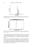
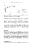
198 JOURNAL OF COSMETIC SCIENCE controls (100 ± 2.7%) however, concentrations of 0.32 and 0.64% (116.3 ± 6.4% and 116. 7 ± 3.3%, respectively) showed a significant effect on fibroblast proliferation com pared to the controls (p 0.05) (Figure 1). Ascorbic acid concentrations of 0.01 and 0.1 mM (110.2 ± 9.4% and 110.6 ± 27.5%, respectively) did not show a significant effect on fibroblast proliferation compared to the controls (100 ± 3.1 %) however, concentrations of 1.0 and 10 mM (120.9 ± 24.1 % and 135.3 ± 33.6%, respectively) showed a signif icant effect on fibroblast proliferation compared to the controls (p 0.05) (Figure 2). EFFECTS OF PLACENTAL EXTRACT AND ASCORBIC ACID ON TGF-131 EXPRESSION The expression of TGF-�1 was determined by ELISA. No significant differences in TGF-�1 expression were observed after treatment with placental extract at concentra tions of 0.08, 0.16, 0.32, and 0.64% (99.9 ± 3.7%, 99.4 ± 7.2%, 100.2 ± 6.4%, and 106.9 ± 0.9%, respectively) compared to the controls (100 ± 4.2%) (p 0.05). More over, TGF-�1 expression did not increase significantly when fibroblasts were treated with a concentration of placental extract greater than 0.64%. Conversely, a significant increase in TGF-� 1 expression occurred after treatment with ascorbic acid at concen trations of 1.0 and 10 mM (107.3 ± 9.7% and 120.33 ± 17.8%, respectively) compared to the controls (100 ± 5.4%) (p 0.05). The results are shown graphically in Figures 3A and 3B. DISCUSSION The activity of human placental extract has become a matter of increasing interest, and 140 Fibroblast Proliferation * * 120 100 - 80 'o 60 40 20 0 control 0.08 0.16 0.32 0.64 Placental extract (1At) Figure 1. The effects of placental extract on fibroblast proliferation. Fibroblasts were treated with placental extract at concentrations of 0.08, 0. 16, 0.32, and 0.64%. Concentrations of 0.32 and 0.64% resulted in significant differences in fibroblast proliferation compared to controls. *Significantly different from controls (p .05).
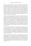
Purchased for the exclusive use of nofirst nolast (unknown)
From: SCC Media Library & Resource Center (library.scconline.org)