226 JOURNAL OF COSMETIC SCIENCE in order to overcome the limitations of STM and broadened the base of applications for SPM. Historically, the field of material science was the first to embrace the technology for nanoscale measurements (1-4). The range of the applications in particulate matter reaches from nanoparticle morphology visualization to quantitative characterization of a single particle to ensemble-like measurements of complex distributions (5). The AFM technique percolated into adjacent fields of colloidal chemistry, biology, biochemistry, and biomedicine. Currently it is being utilized in dentistry and the pharmaceutical industries. Phenomena regarding particle conformation, particle particle interaction, agglomeration, and adhesion have recently attracted the attention of many researchers in these fields (6-8). AFM structural investigations of viruses in solution have brought to light many answers that had been out of reach by other techniques (9). AFM studies of tooth enamel at the nanoscale range has been one focus of interest for dentistry (10,11). The goal of this paper is to show how AFM capabilities that have been traditionally used in material science can now be employed in the cosmetics industry to advance product development. A more comprehensive review of AFM can be found in the literature (3,4,12). In brief, AFM application advantages and capabilities can be summarized as follows: (a) Particles and surfaces can be imaged and characterized quantitatively in ambient conditions, in intervening media including liquids, or in an ultra-high vacuum (UHV). (6) Electric conductivity of the samples is not required and sample preparation is minimal (13). (c) Physical properties of materials, such as magnetic, electrical, thermal, and mechanical parameters, can also be measured. EXPERIMENT AL REPLICA SPECIMEN A negative replica of the skin surface was made using silicon rubber material mixed with a catalyst (Xantropen L Blue with Cuttersil). Replicas overlying the glabella were taken at baseline and repeated after eight weekly AcuFacial treatments at the UCLA Center for East-West Medicine. AcuFacial treatments included acupuncture, Chinese-style facial massage, and patient education on self-acupressure. Lifestyle aspects such as stress man agement, exercise, sleep, and diet were also addressed. Information on the different levels of effectiveness of the various topical prescription and over-the-counter anti-aging prod ucts on the market enabled patients to make informed decisions about the use of these products. For both the baseline and post-treatment replicas, the subject was placed in a supine position. Eyes were closed, and contractions of facial muscles were avoided. A replica ring locator (Replica™ Standard Replica Locator) was placed overlying the glabella, using the root of the nose and eyebrow ridges as landmarks. On a clean tray, the silicon rubber material was mixed together with the catalyst for 15-20 seconds in a ratio of 1 drop of catalyst per 1 ml of rubber material. The 1-2 ml of mixture paste was smeared over the replica to a final thickness of 2-3 mm. After one minute of polymerization, the impression was removed together with the replica ring. Orientation of the replica ring

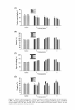
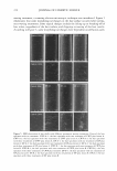
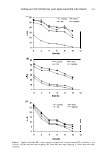
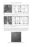
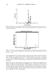
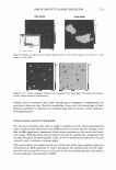
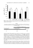
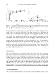
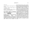
AFM IN PARTICLE CHARACTERIZATION 227 and date were recorded in permanent marker. The replica samples were examined with both SEM and AFM. We used variable-pressure SEM 5136 LSU (Tescan) for the SEM study of the topography of the skin replicas. No special preparation for the replicas was applied. The variable pressure capabilities of this machine precluded specimen coating with conductive materials such as with conventional SEM. Images were taken at 20 kV, and the chamber pressure was maintained at 30 Pa. BI-MODAL SUSPENSION SPECIMEN The polymer nanoparricle mixture was prepared by mixing mono-modal suspensions in a ratio of 1: 1 by volume. NIST-traceable polymer nanospheres (Duke Scientific, Inc) with certified nominal diameters of 102±3 nm and 21 ± 1.5 nm were used. The standard deviation of the distribution for 102-nm spheres was 4.4%. The standard deviation of the distribution for 21-nm spheres was not specified. The AFM specimens were prepared by spin coating the solution on Nanoflat™ substrates and placed in an EClOl spin coater (Headway Research Inc.). Diluted solutions were deposited on the substrate and allowed to sit for 30 seconds prior to spin coating at 2000 rpm for 30 seconds. The solutions were characterized by AFM. The water suspension mixture was characterized by DLS (Wyatt Technologies Inc). All AFM data shown in this paper were obtained using a Light Lever Nano-Rp™ AFM (Pacific Nanotechnology Inc.) and analyzed with NanRule+ TM image analysis software. Nanoflat™ substrates were used for the sample preparation. The vibrating mode or close contact mode with a Si probe (Applied Nanostructure Inc.) was used for imaging. RESULTS AND DISCUSSION APPLICATION TO REPLICA EVALUATION (ANTI-WRINKLE TREATMENT) The example of the 3D visualization of the replica surface roughness is shown in Figure 1. The height profile data (Figures 2 and 3) show that the RMS surface was 48 nm at baseline and 25 nm after eight weeks of treatment. In the case of line-depth measure ments, the depth was 145 nm, 132 nm, and 115 nm at baseline, and the corresponding areas measured 85 nm, 72 nm, and 56 nm after eight weeks of treatment. Both depth and surface roughness were greater at baseline than after treatment. 5"an D.Utance IZ3, UJlldl Z D.Utance (655.ZSIIID! i 655,25m 327,63 ... f Z 0.00 ... ez, DistMC:e (17,11?1 z :01atllft011: (494 .eoma, i 484.80""' 242.40mn o.oo ... Figure 1. 3D AFM image (24 x 24 microns) of pretreatment replica (left). 3D AFM image (17 x 1 7 microns) of posttrearment replica (right).
Purchased for the exclusive use of nofirst nolast (unknown)
From: SCC Media Library & Resource Center (library.scconline.org)