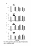
236 JOURNAL OF COSMETIC SCIENCE needle (outer diameter = 0.91 mm) used as a nozzle, and by connecting the grounding electrode to a home-made rotating metal drum, used as the fiber-collecting device. The electric field was fixed at 15 kV/15 cm. For the morphological study, the collection time was about 5 min, while, for the rest of the experiments, the collection time was about 24 h. The drum (outer diameter = 15 cm) rotated at a speed of about 50-65 rpm. The feed rate of the solutions was controlled to about 1 ml h - l using a syringe pump. The GM extract-loaded PVA films were prepared by the solution-casting technique from a PVA solution having a concentration of 10% w/v. The thickness of both the electro spun fiber mats (for the ones that were electrospun for about 24 h) and the cast films was controlled between 20 and 30 µm. CHARACTERIZATION OF NEAT AND GM EXTRACT-LOADED ELECTROSPUN PVA FIBER MATS The morphological appearance of both the neat and the GM extract-loaded electrospun PVA fiber mats was observed by a scanning electron microscope (SEM model S-3400N, Hitachi, Japan). The electrospun fiber mats were sputtered with a thin layer of gold prior to SEM observation. Based on these SEM images, the average diameters of the electro spun fibers could be measured and were reported as average values from at least 100 measurements. The degree of swelling and weight loss of both the neat and the GM extract-loaded electrospun PVA fiber mats was measured in distilled water at room temperature for 24 h according to the following equation: Degree of swelling ( % ) = (M - Md)/Md X 100 (1) where M is the weight of each sample after submersion in water for 24 h and M d is the initial weight of the sample in its dry state. RELEASE OF GM EXTRACT-LOADED ELECTROSPUN PV A FIBER MATS AND CAST PV A FILMS Actual GM extract content. The GM extract content in the GM extract-loaded electrospun PVA fiber mats and cast PVA films was quantified by dissolving the specimens in 4 ml of distilled water for 24 h. After that, 0.5 ml of the solution was pipetted and the GM extract content was measured based on the antioxidant activity. Release of GM extracts. To study the release characteristics of the GM extracts from the GM extract-loaded electrospun PVA fiber mats and as-cast PVA films, a total immersion method was carried out. GM extract-loaded electrospun PVA fiber mat specimens (5 cm2) or cast PVA film specimens (0.5 cm2 ) were immersed in 2 ml of distilled water at 32°C. At a given submersion time point (5, 10, 30, 60, 90, or 120 min), 0.5-ml aliquots of the released medium were withdrawn and the same volume of fresh medium was added. The antioxidant activity in the sample solutions was analyzed. All experiments were performed in triplicate. STATISTICAL ANALYSIS All results were expressed as mean ± S.D. Data were analyzed by one-way analysis of

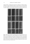
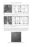
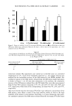
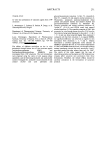
ELECTROSPUN PV A FIBER MATS AS EXTRACT CARRIERS 2 3 7 variance (ANOVA) followed by an LSD post hoc test. Differences of p 0.05 were considered statistically significant. RESULTS AND DISCUSSION MORPHOLOGY OF NEAT AND GM EXTRACT-LOADED ELECTROSPUN PVA FIBER MATS The 10% w/v PV A solution in distilled water was electrospun under an electrical potential of 15 kV applied over a collection distance of 15 cm (i.e., the electric field of 15 kV/15 cm). Figure la demonstrates a selected SEM image of the electrospun PVA fibers. Clearly, cross-sectionally round fibers with a smooth surface were obtained. The average diameter of these fibers was 197 .3 ± 49.8 nm. Based on the good quality of the obtained fibers, 10% w/v PVA solution was then used as the base solution into which the GM extracts were added. After completely dissolving the GM extracts at 2.5%, 5%, and 10% w/w (based on the weight of PV A), the resulting solutions were electrospun. Selected SEM images of the GM extract-loaded electrospun PVA fibers are shown in Figure 1 b-d. The addition of the GM extracts within the base PVA solution did not affect the morphological ap pearance of the obtained fibers, although the fiber diameter tended to decrease as the GM Figure 1. Scanning electron micrograph (10000 x) of electrospun PVA fiber mats. (a) bare electrospun PVA fiber mats. (b) 2.5% initial GM extract-loaded electrospun PVA fiber mats. (c) 5% initial GM extract-loaded electrospun PVA fiber mats. (d) 10% initial GM extract-loaded electrospun PVA fiber mats.
Purchased for the exclusive use of nofirst nolast (unknown)
From: SCC Media Library & Resource Center (library.scconline.org)