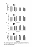
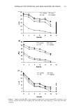
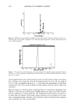
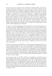
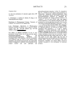
EFFECTS OF PLACENTAL EXTRACT ON FIBROBLAST PROLIFERATION 199 180 Fibroblast Proliferation 160 140 120 100 80 0 0 60 40 20 0 control 0.01 0.1 1 10 L-ascorbic acid (mM) Figure 2. The effects of ascorbic acid on fibroblast proliferation. Fibroblasts were treated with placental extract at concentrations of 0.01, 0.1, 1.0, and 10 mM. Concentrations of 1.0 mM and 10 mM resulted in significant differences in fibroblast proliferation compared to controls. *Significantly different from controls (p .OS). it has been shown to increase peripheral circulation, stimulate cell respiration and tissue metabolism, reduce inflammation, prevent allergies and pigmentation, promote granu lation and removal of old keratin, moisturize, and eliminate activated oxygen. Thus, many dermatologists are interested in its therapeutic properties. Human placenta is an extremely rich reservoir of bioactive molecules. The presence of bioactive peptides in human placenta, such as endothelin (ET)-1 (9,10), adrenocorricotropic hormone (ACTH) (11,12), and sphingolipids (13,14) is well documented (15). ET-1 is a versatile peptide that demonstrates significant mitogenic (16), dendricity-inducing (17,18) and melano genic (19) activity in melanocytes. ACTH has been reported to play an important role in melanogenesis (20). Sphingolipids and their metabolites act as crucial second mes senger molecules that control the rheostatic switch that balances cell growth promotion and inhibition signals (21-23). Human placenta has been used in cosmetics and skin-care soaps due to its anti inflammatory, anti-anaphylactic, antioxidative, anti-melanogenic, melanizing, moistur izing, and collagen-synthesizing properties. In a previous study, we investigated the effects of placental extract on the proliferation and melanogenesis of pigment cells and showed that placental extract may be an effective agent in the treatment of pigment disorders aggravated by ultraviolet light (24). In a separate in vitro study, Sarkar et al. showed that placental protein/peptide fraction-mediated increases in tyrosinase expres sion occurred via transcriptional upregulation, which stimulated melanogenesis in

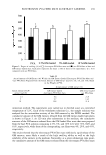
200 JOURNAL OF COSMETIC SCIENCE 140 TGF-ll1 level 120 - c 100 80 C - 60 0 C 40 II 20 0 control 0.08 0.16 0.32 0.64 A Placental extract(%) TGF-131 level * 140 - 120 � 100 C 80 0 0 60 C cu � 40 20 0 control 0.01 0.1 1 10 B L-ascorbic acid (mM) Figure 3. TGF-�1 protein expression determined by ELISA. (A) No significant difference in TGF-�1 expression was observed after treatment with placental extract at all concentrations. (B) A significant increase in TGF-�1 expression was seen after treatment with ascorbic acid at 1.0 and 10 mM compared to controls. *Significantly different from controls (p .05 ). B16F10 cells and primary melanocytes (29). Although the literature describes the anti-melanogenic effects of placental extract, we were not able to find any studies on the effects of placental extract on fibroblast proliferation-associated collagen synthesis. Thus, we compared the effects of placental extract on fibroblast proliferation with those of
Purchased for the exclusive use of nofirst nolast (unknown)
From: SCC Media Library & Resource Center (library.scconline.org)