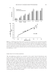
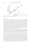
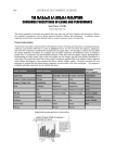
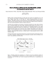
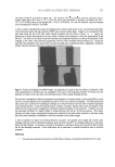
JOURNAL OF COSMETIC SCIENCE 484 these cells at the genomic level. Proof will likely come through more intimate studies done under conditions of hypoxia, where it is known that cellular cytoglobin upregula- tion has been demonstrated. However, even in these studies, separating genetic infl uences from oxygen-shuttling infl uences will remain fundamentally diffi cult. ACKNOWLEDGMENTS Samples of purifi ed human-derived cytoglobin and purifi ed plant-based leghemoglobin were kind gifts of Professor Mark Hargrove, Iowa State University, Ames, Iowa. REFERENCES (1) J. T. Trent and M. S. Hargrove, A ubiquitously expressed human hexacoordinate hemoglobin, J. Biol. Sci., 277, 19538–19545 (2002). (2) T. Burmester, B. Ebner, B. Weich, and T. Hankeln, Cytoglobin: A novel globin type unquitously ex- pressed in vertebrate tissues, Mol. Biol. Evol., 19, 416–421 (2002). (3) US Patent 20030198700A1. (4) M. Schmidt, F. Gerlach, A. Avis, T. Laufs, S. Wystub, J. C. Simpson, E. Nevo, S. Saaler-Reinhardt, S. Reuss, T. Hankeln, and T. Burmeser, Cytoglobin is a respiratory protein in connective tissue and neurons, which is upregulated by hypoxia, J. Biol. Chem., 279, 8063–8069 (2004). (5) K. Nakatani, H. Okuyama, Y. Shimahara, S. Saeki, D. Kim, Y. Nakajima, S. Seki, N. Kawada, and K. Yoshizato, Cytoglobin/STAP, its unique localization in splanchnic fi broblast-like cells and function in organ fi brogenesis, Lab. Invest., 84, 91–101 (2004). (6) A. Pesce, M. Bolognesi, A. Bocedi, P. Ascenzi, S. Dewilde, L. Moens, T. Hankeln, and T. Burmester, Neuro- globin and cytoglobin: Fresh blood for the vertebrate globin family, EMBO Reports, 3, 1146–1151 (2002). (7) D. Hamdane, L. Kiger, S. Dewilde, J. Uzan, T. Burmester, T. Hankeln, L. Moens, and M. C. Marden, Hyperthermal stability of neuroglobin and cytoglobin, FEBS J., 272, 2076–2085 (2005). (8) E. Fordel, E. Geuens, S. Dewilde, P. Rottiers, P. Carmeliet, J. Grooten, and L. Moens, Cytoglobin ex- pression is upregulated in all tissues upon hypoxia: An in vitro and in vivo study by quantitative real- time PCR, Biochem. Biophys. Res. Commun., 319, 342–348 (2004). (9) P. P. A. Mammen, J. M. Shelton, Q. Ye, S. B. Kanatous, A. J. McGrath, J. A. Richardson, and D. J. Garry, Cytoglobin is a stress-responsive hemoprotein expressed in the developing and adult brain, J. Histochem. Cytochem., 54, 1349–1361 (2006). (10) X. Guo, S. Philipson, and K. Tan-Un, Study of the hypoxia-dependent regulation of human CYGB gene, Biochem. Biophy. Res. Commun., 364, 145–150 (2007). (11) D. Li, X. Q. Chen, W. Li, Y. Yang, J. Wang, and A. C. H. Yu, Cytoglobin up-regulation by hydrogen peroxide plays a protective role in oxidative stress, Neurochem. Res., 32, 1375–1380 (2007). (12) E. Fordel, L. Thijs, L. Moens, and S. Dewilde, Neuroglobin and cytoglobin expression in mice: Evidence for a correlation with reactive oxygen species scavenging, FEBS J., 274, 1312–1317 (2007). (13) N. J. Hodges, N. Innocent, S. Dhanda, and M. Graham, Cellular protection from oxidative DNA dam- age by over-expression of the novel globin cytoglobin in vitro, Mutagenesis, 23, 293–298 (2008). (14) E. Geuens, I. Brouns, D. Flamez, S. Dewilde, J. Timmermans, and L. Moens. A globin in the nucleus! J. Biol. Chem., 278, 30417–30420. (15) A. Fago, C. Hundahl, H. Malte, and R. E. Weber, Functional properties of neuroglobin and cytoglobin. Insights into the ancestral physiological roles of globins, IUBMB Life, 56, 689–696 (2004). (16) M. M. Stevens and J. H. George, Exploring and engineering the cell surface interface, Science, 310, 1135–1138 (2005). (17) J. V. Gruber, Globin proteins: A fi rst line of defense against reactive oxygen species, Cosmet. Sci. Technol., 1, 69–75 (2005). (18) C. Seregelyes and D. Dudits, Phytoglobins and nitric oxide: New partners in an old signaling system in plants, Acta Biol. Hung., 54, 15–25 (2003). (19) P. K. Muller, E. Kirsch, V. Gauss-Muller, and T. Krieg. Some aspects of the modulation and regulation of collagen synthesis in vitro, Mol. Chem. Biochem., 28, 73–85 (1981).

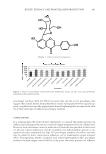
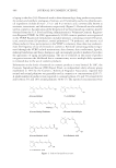
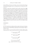
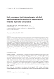
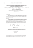
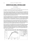
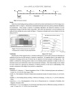
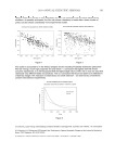
J. Cosmet. Sci., 60, 485–500 (September/October 2009) 485 Determination of retinol, retinyl palmitate, and retinoic acid in consumer cosmetic products JEAN C. HUBINGER, U.S. Food and Drug Administration, 5100 Paint Branch Parkway, College Park, MD. Accepted for publication March 26, 2009. Synopsis Retinol and retinyl palmitate are frequently used in cosmetic products. A simple, rapid, and sensitive re- versed-phase high-performance liquid chromatography (HPLC) method with ultraviolet (UV) detection was developed for the quantitation of retinol, retinyl palmitate, and retinoic acid in cosmetic preparations. The analytes were extracted from a cosmetic/Celite mixture using a solvent system composed of equal amounts of hexane, isopropanol, and ethyl acetate, and the extract was injected directly into an HPLC chromatograph with a C18 column and UV detector set at 330 nm. Chromatographic separation was achieved by gradient elution with a mobile phase, starting with aqueous ammonium acetate buffer/methanol that was gradually changed to methanol/dichloromethane. The average recoveries of retinol, retinyl palmitate, and retinoic acid from spiked cosmetic products were 95% or higher. In a survey of twenty-nine consumer cosmetic skin care products labeled to contain retinoids, most products were found to contain either retinol or retinyl palmitate at concentrations up to 2.2% (w/w), while a few products contained both ingredients. A number of products also contained cis isomers of retinol that could be quantitatively distinguished from the all-trans compound. The method can be used to quantitate several retinoids and their isomers in cosmetic products. The method will be useful for obtaining information needed to estimate levels of exposure to retinoids from cosmetic products. INTRODUCTION Retinol (vitamin A) is the parent compound of a large number of natural and synthetic compounds collectively referred to as retinoids. In addition to retinol, the primary bio- logically important and naturally occurring retinoids are retinyl palmitate, retinalde- hyde, and retinoic acid (Figure 1). These retinoids are essential for the development, growth, and health of vertebrates. One of the earliest biological functions discovered for retinoids was their critical role in the development and maintenance of healthy epithelial tissue, including the skin (1–3). Studies have demonstrated that the normal structure and function of the skin is dependent on the orchestration of cellular division, differentiation, and keratinization by retinoids (4). Discovery of the powerful effects elicited by retinoids in the skin has led to their wide- spread use in dermatologic drug products and cosmetics. Both topically and orally ad- ministered retinoids are currently used to treat dermatologic conditions such as acne and disorders of keratinization (e.g., psoriasis and ichthyosis) and to reduce the clinical signs
Purchased for the exclusive use of nofirst nolast (unknown)
From: SCC Media Library & Resource Center (library.scconline.org)