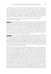
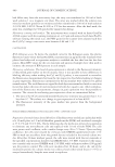
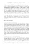
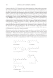
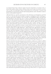
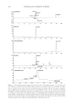
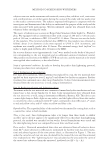
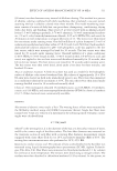
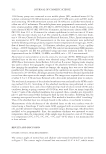
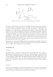
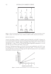
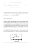
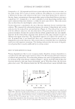
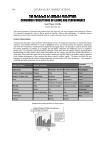
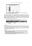
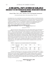
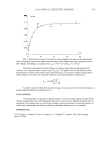
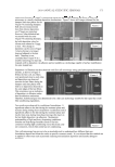
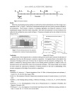
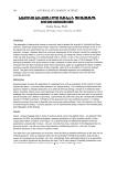
METHOD TO SCREEN SKIN WHITENERS 503 subjects were not under treatment with retinoids, tetracycline, nalidixic acid, corticoster- oids, antihistamines, or similar agents during the course of the study and two weeks prior to the study’s commencement. The subjects expressed willingness to cooperate with the investigator and demonstrated the ability to understand the purpose of the study and the risks associated with participation. Written informed consent was obtained from each volunteer before entering the study. The source of radiation was a xenon arc Berger Solar Simulator (Solar Light Co., Philadel- phia, PA) equipped with an interference fi lter with a range of 280 nm to 320 nm and a peak of 300 nm, in addition to WG 320 and UG-11 fi lters. The test site was the backs of the panelists. The minimal erythemal dose (MED) for the panelists was obtained as follows: about 2-cm diameter circles were exposed to UV-B in 25% increments, and erythema was visually graded after 24 hours. The minimal energy level (mJ/cm2) to induce a slight pink erythema after 24 hours is the MED. Six to seven distinct areas (approximately 4 cm2) were marked on the backs of the panel- ists corresponding to the test materials and an additional untreated irradiated control. The panelists received twice the MED of UV-B on each site, and the materials to be tested were applied after irradiation, as described below. Setup of experimental conditions. In order to develop the perfect skin lightening protocol, various procedures were investigated: Part I: Evaluation of tan reduction Five days after irradiation, when erythema was replaced by a tan, the test materials were applied on their respective sites (2 mg/cm2) and allowed to dry for ten minutes. Product treatment was continued once a day except Sunday for 21 days. Color measurements were obtained from the test sites twice a week, using a chromameter. Part II: Reduction of onset and lightening of tan Treatment with the test materials was commenced immediately after irradiation and con- tinued once a day, except Sunday, for 21 days. Color measurements were obtained from the test sites twice a week, using a chromameter (Minolta, Ramsey, NJ). This device uses a xenon light lamp to fl ash a light on any surface, and the light refl ected from this surface is converted to color co-ordinates where L* values correspond to skin refl ectance, a* values to red and yellow color, and b* values to yellow and blue color. Reproducibility. The reproducibility of the method was determined by testing kojic acid as a control fi ve times in both methods. Three vs four weeks. Since hydroquinone takes a lot longer than three weeks to exhibit an effect and it did not appear to be signifi cantly effective in this short skin-lightening protocol, the method was extended to four weeks. Several formulations were tested and the skin-lightening factor was compared between three and four weeks. Data analysis. Untreated unirradiated skin color was subtracted from all values to deter- mine ΔL* values (decrease in refl ectance), normalized for baseline (one day after irradia- tion) and plotted against all the time points (days), as illustrated in Figure 1. The area under the curve for each test site was calculated for the treated (At) site and the untreated (Ac) irradiated site. The “lightening factor” was calculated as the area under the curve of the treated site subtracted from the untreated, irradiated site (LF = Ac − At).

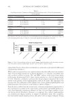
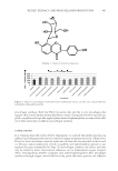
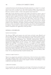
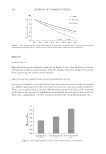
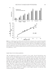
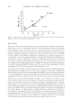
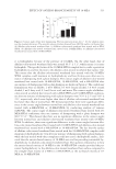
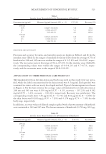
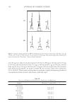
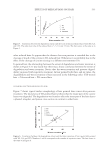
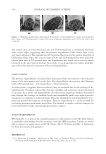
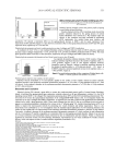
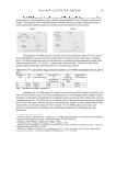
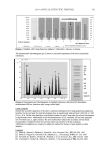
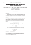
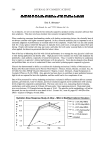
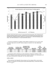
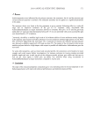
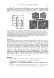
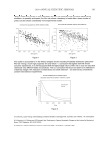
JOURNAL OF COSMETIC SCIENCE 504 RESULTS REPRODUCIBILITY Reproducibility of the method is exhibited in Figure 2. It is clear that kojic acid gave reproducible results in both methods, with the standard deviation within 2–5% of the mean, indicating the validity of this method. REDUCTION OF TAN VS REDUCTION OF ONSET AND INTENSITY OF TAN As observed in Figure 3a, the lightening of the onset and the intensity of the tan resulted in a higher lightening factor than the reduction of the tan that was already established. This is to be expected since the anti-infl ammatory properties of some of the materials would reduce the intensity of infl ammation and thereby the intensity of the initial tan. There was a signifi cant (p 0.001) correlation between the two methods (Figure 3b). Figure 1. Skin lightening after three and four weeks of treatment. Area under the curve of untreated–treated exhibited the lightening factor, which can be calculated after three- and four-week treatments. Figure 2. Skin-lightening effect of kojic acid and repeatability of the methods.
Purchased for the exclusive use of nofirst nolast (unknown)
From: SCC Media Library & Resource Center (library.scconline.org)