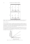
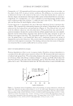
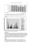
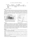
J. Cosmet. Sci., 60, 501–508 (September/October 2009) 501 A novel method to study the skin-lightening effect of topical materials NEELAM MUIZZUDDIN, KENNETH D. MARENUS, THOMAS MAMMONE, and DANIEL H. MAES, Estee Lauder Companies, Melville, NY 11747. Accepted for publication April 1, 2009. Synopsis Skin without signifi cant dyschromia is an aesthetic requirement for people worldwide. There are several in vitro methods to determine the whitening potential of actives however, the in vivo testing of skin whiteners is a long and expensive process. We have designed a rapid clinical method to screen potential skin whiteners using a UV-induced skin tan as a model. Small areas of identical suntan are repeatably induced on the skin, and treatment of these sites allows rapid screening of several skin whiteners within the course of a month. The method provides reproducible results and valuable information about the potential skin-lightening activity of topical preparations. INTRODUCTION In Asia, Africa, and South America, the popularity of fair-skinned beauties in the media, as well as the cultural preference towards lighter skin, has fuelled public demand for skin- lightening products (1). The tendency of pigmented skin to be more prone to develop hyperpigmentation has also contributed to this demand (2). In most instances, sun exposure is the main stimulator of skin hyperpigmentation. A direct effect of UV photons results in activation of tyrosinase, the rate-limiting enzyme in melanin synthesis, as well as an increase in cell surface expression of receptors for at least one of the several known keratinocyte-derived melanogenic factors, MSH (3). The inherent defense mechanism of skin against UV-B allows the transformation of tyrosine to 3,4-dihydroxyphenylalanine in the presence of UV-activated tyrosinase. This is further oxidized to DOPA quinone and then to dopachrome (4). Reduction of dopachrome yields 5,6-dihydroxyindole-2-carboxylic acid, which is ultimately converted via dihydroxyin- dole to the yellow-colored indole 5,6-quinone. Further, oxidative coupling of this results in the formation of eumelanin. Under normal circumstances eumelanins and pheomela- nins are formed simultaneously to lead to the so-called mixed-type melanins. This process requires three to six days before the tan is established (4). There is a myriad of skin whiteners with variable effi cacy (5) readily available for consum- ers. Modulation of melanogenesis in the melanocytes can be achieved using chemicals that share structural homologies with the substrate tyrosine and as thus competitively

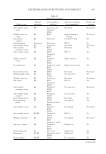
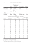
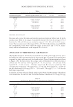
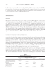
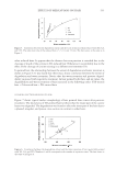
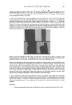
JOURNAL OF COSMETIC SCIENCE 502 inhibit the catalytic function of tyrosinase (6). Kojic acid inhibits the catecholase activity of tyrosinase, which is the rate-limiting, essential enzyme in the biosynthesis of the skin pigment melanin. Hydroquinone, a hydroxyphenolic chemical, is a bleaching agent that has been used for decades as a skin-lightening agent (7). It acts by inhibiting the enzyme tyrosinase, thereby reducing the conversion of DOPA to melanin. Some of the other mechanisms of action are the destruction of melanocytes, degradation of melanosomes, and the inhibition of the synthesis of DNA and RNA (8). Strong skin lighteners like clobetasol and hydroquinone can cause complications and signifi cant health problems, especially for individuals with skin of color (9–11). There- fore, there is a constant need to investigate new and diverse potential skin lighteners that are safer and do not have side effects. Determination of the skin-whitening abilities of actives requires three-to-six-month, in- use, clinical studies. Several techniques are used to assess skin lightening such as clinical inspection, photographs, or the use of surface color measuring devices such as a chro- mameter. Photographs are obtained at various time points, followed by image analysis (12) and/or visual analysis (13,14). Chromameters have been used to determine skin color before and after treatment, but these measurements require strict controls to account for the suntan during the course of the study. Most of these studies require a large number of subjects and a commitment of several months to screen a sample along with its controls. We developed a simple and quick method to screen several materials for skin lightening that can be carried out within a month. MATERIALS AND METHODS MATERIALS Several actives were tested including chamomile 1% (E.L. Japan), magnesium ascorbyl phosphate 0.1% (Presperse), hydroquinone 2% (Sigma), and kojic acid 2% as standard (Sigma). All these actives were tested in a silicone-in-water formulation base, which was tested by itself as the placebo (vehicle). In addition, several commercial lightening formulations were tested. These formulations contained various skin lighteners and were coded as whitening mix 1, 2, and so on. Tri- luma (Galderma) containing 4% hydroquinine was also tested. Triluma also contains fl uocinolone acetonide, an anti-infl ammatory corticosteroid and tretinoin. In order to determine if exfoliants would have an effect of skin lightening in this protocol, a formula- tion containing 1% salicylic acid was also tested. PROCEDURE The study was conducted at a contract testing laboratory in New City, New York. In each test, eight to ten subjects were recruited from the local population. Males or females ages 21–48 with no evidence of acute or chronic disease, including dermatological or ophthal- mologic problems, were enrolled in the study. In order to qualify, the Caucasian subjects were required to be skin type III, who tan readily. The skin of the back was required to be free of warts, nevi, moles, sunburn, suntan, scars, and active dermal lesions. The
Purchased for the exclusive use of nofirst nolast (unknown)
From: SCC Media Library & Resource Center (library.scconline.org)