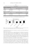
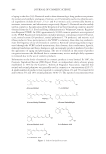
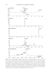
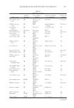
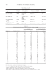
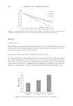
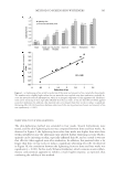
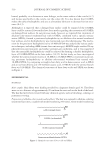
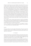
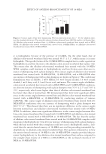
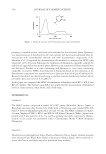
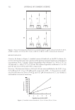
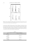
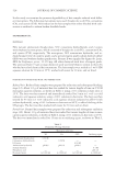
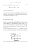
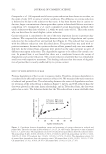
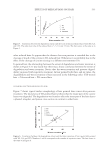
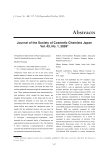
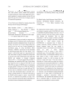
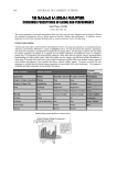
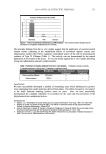
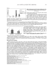
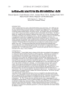
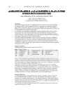
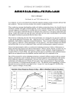
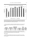
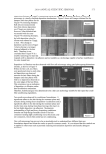
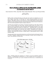
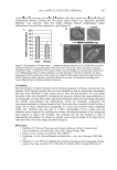
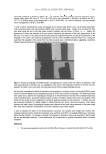
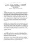
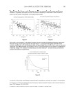
METHOD TO SCREEN SKIN WHITENERS 505 THREE-WEEK VS FOUR-WEEK LIGHTENING The skin-lightening method was extended to four weeks. Several formulations were tested, and the skin-lightening factor was compared between three and four weeks. As observed in Figure 4, the lightening factor after four weeks was higher than after three weeks probably because the additional time allowed further whitening activity. Hydro- quinone can be irritating on skin, especially infl amed skin (8), and as a result it was not that effective when applied soon after irradiation. In addition, this material takes a lot longer than three to four weeks to induce a signifi cant whitening effect (8). As observed in Figure 3b, the correlation between the lightening factor at three and four weeks was signifi cant (p 0.001). In this study Triluma (Galderma), which contains an anti-infl am- matory corticosteroid, appeared to exhibit a much higher response, as expected, thus confi rming the validity of this method. Figure 3. (a) Lightening of tan as well as reduction of the onset and intensity of tan, tested after three weeks. The numbers were slightly higher when the test materials were applied soon after irradiation, probably be- cause the materials reduced infl ammation, which is an integral component of skin pigmentation. Hydroqui- none can be irritating on skin, especially infl amed skin (8) as a result, it was not that effective when applied soon after irradiation. In addition, this material takes a lot longer than three weeks to induce a signifi cant whitening effect (8). (b) Correlation between reduction of skin tan vs prevention of onset and intensity of tan was signifi cant (p 0.001).

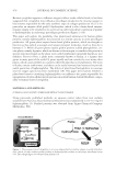
JOURNAL OF COSMETIC SCIENCE 506 DISCUSSION The actual color of skin is determined by the type and amount of melanin synthesized by melanocytes and by its distribution pattern in the surrounding keratinocytes. Melanin forms through a series of oxidative reactions involving the amino acid tyrosine in the presence of the enzyme tyrosinase. Tyrosinase catalyses three different reactions in the bio- synthetic pathway of melanin in melanocytes: the hydroxylation of tyrosine to L-DOPA and the oxidation of L-DOPA to dopaquinone furthermore, in humans, dopaquinone is converted by a series of complex reactions to melanin (15). Production of melanin can sometimes be excessive and uneven, resulting in a dark and discolored skin tone, which is a serious concern in health and beauty maintenance. As a result there is a myriad of skin-lightening and depigmenting products available, which contain actives like magnesium- 1-ascorbyl-2-phosphate (MAP), hydroxyanisole, N-acetyl-4-S-cysteaminylphenol, arbutin (hydroquinone-beta-d-glucopyranoside), and hydroquinone (15). A literature review shows a variety of methods for testing the effi cacy of whitening agents, most of which are confi ned to in vitro tests in cell cultures (16–18). These methods deal with blocking various steps of the melanogenesis pathway, such as tyrosinase inhibition (17,19) and inhibition of melanosome transfer (20). These methods are valuable for screen- ing purposes but do not translate to how the materials will act on skin in clinical settings. There are also several clinical methods available, most involving 8–12-week, in use, clinical trials in which skin color is graded via visual and instrumental assessments (6). Other clinical studies involve the recruitment of subjects with pigmentation disorders like melasma (21), solar lentigos (6), and post-infl ammatory hyperpigmentation (22). In these studies, photographs of the lesions are obtained before and after several weeks of treatment, followed by image analysis of the photographs to determine the lightening of the lesions. Fluorescence photography is another noninvasive method that is sensitive in the evaluation and quantifi cation of the distribution and changes of mottled and diffuse hyperpigmentation (23). In addition, the overall skin color is assessed by the subjects and trained technicians. All these current methods are valuable and effective, but extremely time-consuming, mainly because whitening actives take a long time to act. Figure 4. Lightening factor after four weeks was higher than after three weeks. Correlation between lighten- ing factor at three and four weeks was signifi cant (p 0.001).
Purchased for the exclusive use of nofirst nolast (unknown)
From: SCC Media Library & Resource Center (library.scconline.org)