KUDZU EXTRACT AND PROCOLLAGEN PRODUCTION 479 at 10,000 rpm for one minute. After the fl ow-through was discarded, the cartridge was spun one fi nal time to remove traces of the wash buffer. The cartridge was transferred to a new collection tube and 25 μl of prewarmed elution solution was added to the cartridge. The cartridge was incubated for two minutes at room temperature and then aRNA was eluted by centrifuging for one minute at 10,000 rpm. This elution was performed one additional time to give a total volume of 45–50 μl of aRNA solution. The fi nal concentra- tion of the aRNA was determined by the Ribogreen assay described above. In addition, the quality of the aRNA was checked via gel electrophoresis as described below. Labeling of aRNA with fl uorescent dyes (Perkin Elmer ASAP RNA Labeling Kit) and purifi cation of labeled aRNA. Labeling: Two tubes were prepared for the labeling process, one for Cy3 labeling (green) and one for Cy5 labeling (red). To the Cy3 tube was added 2 μg of aRNA prepared from the untreated control sample, and enough DEPC·H2O was added to bring the total vol- ume up to about 4 μl. To the Cy5 tube was added 2 μg of aRNA prepared from the sample treated with the test material, and enough DEPC·H2O was added to bring the total volume up to 4 μl. To both tubes was added 5 μl of ASAP labeling buffer and 1 μl of the specifi c dye for each tube (Cy3 or Cy5). The tubes were incubated for 15 minutes at 85 ± 2°C. At the end of the 15-minute incubation period, the tubes were placed on ice to cool and then 2.5 μl of ASAP stop solution was added to each tube. The proportions provided enough sample to run one microarray chip. Purifi cation: To purify the labeled aRNA, a Microcon YM-30 fi lter column (Millipore) was inserted into a collection tube fi lled with 400 μl of TE buffer. The Cy3 and Cy5 probes were combined (12.5 μl of each) and then added to the Microcon fi lter the mix- ture was thoroughly mixed with the TE buffer. The mixture was centrifuged at 12,000 rpm for eight minutes and the fl ow-through was discarded. The column was then washed twice with 400 μl of TE buffer and the fl ow-through was again discarded. After the fi nal wash, the fi lter column was inverted, placed into a new collection tube, and centrifuged at 12,000 rpm for two minutes to collect the probe. The probe was further diluted to a volume of 2–30 μl with residual TE buffer. Microarray hybridization and washing (Agilent Technologies Microarrays). For hybridization, 45 μl of 10X control target RNA (Agilent Technologies In Situ Hybridization Kit) was mixed with 160 μl of DEPC water and 9 μl of 25X Agilent fragmentation buffer. This mixture was incubated at 60°C for approximately 30 minutes in a hybridization oven. At the end of the incubation period, 225 μl of Agilent hybridization buffer was added, along with the fl uorescent aRNA probes prepared above. During the incubation period, an Agilent SUREHYB hybridization chamber was prepared by inserting a glass gasket slide into the bottom half of the chamber. At the end of the incubation period, the hybridiza- tion mixture (approximately 450 μl) was applied to the glass gasket slide and an Agilent Human 1A Oligo microarray chip was placed face down on top of the gasket in such a way that the hybridization solution was sandwiched between the glass gasket slide and the microarray face of the chip. The top half of the chamber was then attached and the connecting thumbscrew tightened. After verifying that there was good bubble formation in the chamber, it was placed into the hybridization chamber for approximately 17 hours at 60°C and rotated at 4 rpm). At the end of the hybridization period, the microarray glass gasket assembly was removed from the SUREHYB chamber and placed in 50 ml of wash solution 1 (6X SSC, 0.005% Triton X-102) at room temperature. After the gasket

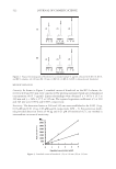
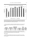
JOURNAL OF COSMETIC SCIENCE 480 had fallen away from the microarray chip, the array was transferred to 300 ml of fresh wash solution 1 on a magnetic stir plate. The array was washed while the solution was mixed at medium speed for ten minutes and then transferred to 300 ml of wash solution 2 (0.1X SSX, 0.005% Triton X-102) at 4°C for fi ve minutes. After the fi nal wash, the array was centrifuged at 500 rpm for fi ve minutes for drying. Microarray scanning and analysis. The microarrays were scanned with an Axon GenePix 4100A scanner with the scanning resolution set at 10 μm and analyzed with GenePix Pro software. During the initial scan, the PMT gains for the scanner were adjusted such that the Cy5/Cy3 image count ratios were between 0.88 and 1.12. CALCULATIONS RNA Ribogreen assay. To derive the standard curve for the Ribogreen assay, the relative fl uorescent units versus the known RNA concentrations in μg/ml for the standards were plotted and subjected to regression analysis to establish the line that best fi ts the data points. Mean RFU values for the test materials and untreated samples were then used to estimate the amount of RNA present in each sample. Microarray calculations. The level of gene expression is related to the fl uorescent intensity of the probed gene marker on the microarray. Since it was possible to have differences in labeling effi ciency when making the Cy3 and Cy5 probes, it was essential to normalize the fl uorescence measurements between the two respective dyes before looking at changes in gene expression. Fluorescence intensities for the microarrays were subjected to global normalization. The total fl uorescent signal for both dyes was normalized with a correction factor that makes the ratio of total intensities for both dyes equal to one. After normaliza- tion of the fl uorescence measurements, changes in gene expression were then possible to examine. The criteria for evaluating gene expression values are summarized below: 1. The ratio of Cy3/Cy5 (untreated/treated) fl uorescence intensity was greater than 1.3 or less than 0.7. This relates to a change in gene expression of at least ±30%. 2. The fl uorescent intensity of the gene marker was greater than the background intensity. HUMAN DERMAL FIBROBLAST PROCOLLAGEN (TYPE 1 C-PEPTIDE) ASSAY Preparation of normal human dermal fi broblasts. Fibroblasts were seeded into individual wells of a 12-well plate in 1.0 ml of fi broblast growth media (FGM) and incubated overnight at 37 ± 2°C and 5 ± 1% CO2. On the following day, the media was removed via aspiration to eliminate any non-adherent cells and replaced with 1.0 ml of fresh FGM. The cells were grown until confl uent, with a media change every 48 to 72 hours. Upon reaching confl uence, the cells were treated for 24 hours with DMEM supplemented with 1.5% FBS to wash out any effects from the growth factors included in the normal culture me- dia. After this 24-hour washout period, the cells were treated with the test materials at the specifi ed concentrations dissolved in DMEM with 1.5% FBS. Untreated cells (nega- tive controls) only received DMEM with 1.5% FBS, while sodium ascorbate (100 μg/ml) was the positive control. The cells were incubated for 48 hours, and at the end of the incubation period, the culture media was collected and either stored frozen (−75°C) or assayed immediately. Samples were tested in triplicate.
Purchased for the exclusive use of nofirst nolast (unknown)
From: SCC Media Library & Resource Center (library.scconline.org)