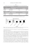
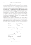
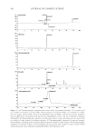
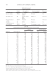
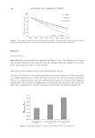
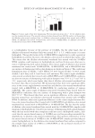
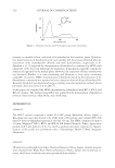
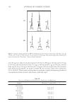
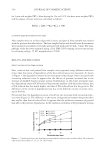
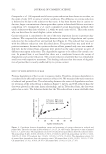
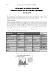
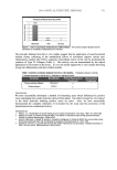
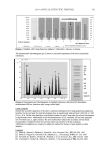
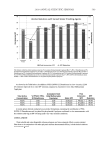
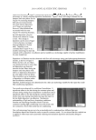
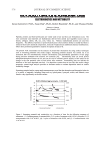
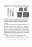
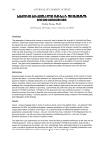
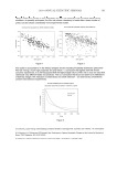
JOURNAL OF COSMETIC SCIENCE 506 DISCUSSION The actual color of skin is determined by the type and amount of melanin synthesized by melanocytes and by its distribution pattern in the surrounding keratinocytes. Melanin forms through a series of oxidative reactions involving the amino acid tyrosine in the presence of the enzyme tyrosinase. Tyrosinase catalyses three different reactions in the bio- synthetic pathway of melanin in melanocytes: the hydroxylation of tyrosine to L-DOPA and the oxidation of L-DOPA to dopaquinone furthermore, in humans, dopaquinone is converted by a series of complex reactions to melanin (15). Production of melanin can sometimes be excessive and uneven, resulting in a dark and discolored skin tone, which is a serious concern in health and beauty maintenance. As a result there is a myriad of skin-lightening and depigmenting products available, which contain actives like magnesium- 1-ascorbyl-2-phosphate (MAP), hydroxyanisole, N-acetyl-4-S-cysteaminylphenol, arbutin (hydroquinone-beta-d-glucopyranoside), and hydroquinone (15). A literature review shows a variety of methods for testing the effi cacy of whitening agents, most of which are confi ned to in vitro tests in cell cultures (16–18). These methods deal with blocking various steps of the melanogenesis pathway, such as tyrosinase inhibition (17,19) and inhibition of melanosome transfer (20). These methods are valuable for screen- ing purposes but do not translate to how the materials will act on skin in clinical settings. There are also several clinical methods available, most involving 8–12-week, in use, clinical trials in which skin color is graded via visual and instrumental assessments (6). Other clinical studies involve the recruitment of subjects with pigmentation disorders like melasma (21), solar lentigos (6), and post-infl ammatory hyperpigmentation (22). In these studies, photographs of the lesions are obtained before and after several weeks of treatment, followed by image analysis of the photographs to determine the lightening of the lesions. Fluorescence photography is another noninvasive method that is sensitive in the evaluation and quantifi cation of the distribution and changes of mottled and diffuse hyperpigmentation (23). In addition, the overall skin color is assessed by the subjects and trained technicians. All these current methods are valuable and effective, but extremely time-consuming, mainly because whitening actives take a long time to act. Figure 4. Lightening factor after four weeks was higher than after three weeks. Correlation between lighten- ing factor at three and four weeks was signifi cant (p 0.001).

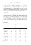
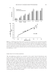
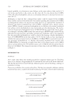
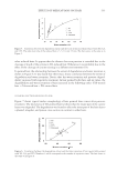
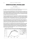
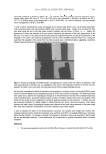
METHOD TO SCREEN SKIN WHITENERS 507 We have developed quick and simple methods to determine the skin-lightening effect of actives, with various modifi cations, depending on the objective of the study. If the test material is designed to reduce pre-existing color, the fi rst method, which addresses reduction of tan, can be employed. If the test material contains anti-infl ammatories and is designed to reduce the onset and intensity of tan, then the second method can be em- ployed. Both these methods involve the use of UV-B induced tanning as the marker. The effect of overexposure to solar ultraviolet radiation (UVR) on human skin has been well described (24,25). The erythema produced is commonly referred to as “sunburn.” UV irradiation initially elicits an infl ammatory reaction (sunburn) that resolves within a few days and converts to a suntan (26). The UV-induced tanning starts to resolve quickly and, depending on the intensity of UV exposure and skin type, is almost gone in four to fi ve weeks. Small areas of identical suntan can be repeatably induced on the skin, and treatment of these sites allows a rapid screening of several skin whiteners within the course of a month. In addition, the lightening factor described in this paper takes into account the whole picture of skin color reduction over the course of the study rather than at individual time points (Figure 1). This method addresses one aspect of skin lightening and by no means refl ects the effect of whiteners on lentigos, melasma, and other skin discolorations. However, it is a valuable tool for screening and choosing the best of several materials and concentrations, and must be followed by the 8–12-week, in-use, clinical trials currently in use. REFERENCES (1) S. B. Adebajo, An epidemiological survey of the use of cosmetic skin lightening cosmetics among traders in Lagos, Nigeria, West Africa, J. Med., 21(1), 51–55 (2002). (2) S. Badreshia-Bansal and Z. D. Draelos. Insight into skin lightening cosmeceuticals for women of color, J. Drugs Dermatol., 6(1), 32–39 (2007). (3) B. A. Gilchrest, H. Y. Park, M. S. Eller, and M. Yaar, Mechanisms of ultraviolet light-induced pigmenta- tion, Photochem. Photobiol., 63(1), 1–10 (1996). (4) F. Ryckmanns, C. Schmoeckel, G. Plewig, and O. Braun-Falco, Early persistent UVA-pigmentation: Ultrastructural and morphometric analyses, Arch. Dermatol. Res., 279(3), 173–179 (1987). (5) J. F. Hermanns, L. Petit, C. Piérard-Franchimont, P. Paquet, and G. E. Piérard, Assessment of topical hypopigmenting agents on solar lentigines of Asian women, Dermatology, 204(4), 281–286 (2002). (6) R. E. Boissy, M. Visscher, and M. A. DeLong, DeoxyArbutin: A novel reversible tyrosinase inhibitor with effective in vivo skin lightening potency, Exp. Dermatol., 14(8), 601–608 (2005). (7) J. L. O’Donoghue, Hydroquinone and its analogues in dermatology—A risk-benefi t viewpoint, J. Cos- met. Dermatol., 5(3), 196–203 (2006). (8) R. M. Halder and G. M. Richards, Topical agents used in the management of hyperpigmentation, Skin Ther. Lett., 9(6), 1–3 (2004). (9) L. Petit, C. Cohen-Ludmann, P. Clevenbergh, J. F. Bergmann, and L. Dubertret, Skin lightening and its complications among African people living in Paris, J. Am. Acad. Dermatol., 55(5), 873–878 (2006). (10) P. Del Giudice, E. Raynaud, and A. Mahé, Cosmetic use of skin depigmentation products in Africa, Bull. Soc. Pathol. Exot., 96(5), 389–393 (2003). (11) R. A. Hoshaw, K. G. Zimmerman, and A. Menter, Ochronosislike pigmentation from hydroquinone bleaching creams in American blacks, Arch. Dermatol., 121(1), 105–108 (1985). (12) J. F. Hermanns, L. Petit, O. Martalo, C. Piérard-Franchimont, G. Cauwenbergh, and G. E. Piérard, Unraveling the patterns of subclinical pheomelanin-enriched facial hyperpigmentation: Effect of depig- menting agents, Dermatology, 201(2), 118–122 (2000). (13) T. Hakozaki, L. Minwalla, J. Zhuang, M. Chhoa, A. Matsubara, K. Miyamoto, A. Greatens, G. G. Hillebrand, D. L. Bissett, and R. E. Boissy, The effect of niacinamide on reducing cutaneous pigmenta- tion and suppression of melanosome transfer, Br. J. Dermatol.,147(1), 20–31 (2002).
Purchased for the exclusive use of nofirst nolast (unknown)
From: SCC Media Library & Resource Center (library.scconline.org)