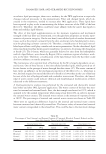
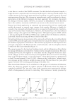
JOURNAL OF COSMETIC SCIENCE 536 The patterns of use for these products are dramatically changing. One notable change is the popularity of tattoos and permanent makeup. It has been reported that 24% of the population in the contiguous United States have at least one tattoo (6). The highest inci- dence of tattoos (36%) occurred for individuals aged 18 to 29. Another change in the pattern of use is the formulation of inks with novel pigments, which allows a wide range of colors for tattoos and permanent makeup. Increasingly, inks are being formulated with organic pigments that have a limited history of safe use (7–9). The high prevalence of tattoos and permanent makeup combined with the use of novel pigments increases the risk for adverse reactions to these products. As an example of this increased risk, the FDA had received only fi ve reports of adverse reactions after permanent makeup procedures between 1988 and 2003 (10). In contrast, between 2003 and 2004, the FDA received 150 reports of adverse reactions attributable to use of inks traceable to one manufacturer of permanent makeup inks. The FDA warned consumers about the adverse reaction re- ports, and the inks were subsequently voluntarily withdrawn from the market (10,11). Reliable toxicological testing methods are needed to assure the safety of tattoo and per- manent makeup inks. However, there have been very few attempts to develop in vitro methods for identifying hazardous inks. Falconi et al., (12) have used human fi broblasts derived from gingival tissue to investigate the cytotoxicity of a permanent makeup ink. The cytotoxicity, resulting from exposures to the ink for up to two weeks, was assessed by measuring inhibition of the fi broblasts’ capacity to metabolize 3-(4,5-dimethylthiazol-2- yl)-2,5-diphenyltetrazolium bromide (MTT assay). Dose-dependent cytotoxicity was ob- served for the selected ink. We have developed an in vitro assay that allows evaluation of both the cytotoxicity and photocytotoxity of inks. Because inks used for decorative tat- toos and permanent makeup contain pigments that are effi cient chromophores and these inks are applied to sun-exposed skin, phototoxicity is a concern. There have been reports of phototoxic reactions resulting from decorative tattoos and permanent makeup (13–20). Although the in vitro assay described here was developed to assess the cytotoxicity and photocytotoxicity of permanent makeup inks, the method is generally more applicable for testing inks used for either decorative tattoos or permanent makeup. In our initial studies, we examined the cytotoxicity and photocytotoxicity of permanent makeup inks containing TiO2. These inks were selected for several reasons. First, TiO2 is one of the most widely used pigments in tattoo and permanent makeup inks. Due to its high refractive index, TiO2 frequently functions as an opacifi er in inks (1). Titanium dioxide is also widely used in pigment mixtures to formulate light shades of inks. In addi- tion, the photocatalytic activity of TiO2 has been widely studied. Therefore, TiO2 is an ideal pigment to investigate in the development of an in vitro assay for photocytotoxicity. Finally, there have been reports associating the presence of TiO2 in inks with adverse reac- tions (21,22). Therefore, toxicological data for inks containing TiO2 are needed. EXPERIMENTAL MATERIALS Titanium dioxide (anatase) was purchased from Sigma-Aldrich (Milwaukee, WI). Sam- ples of TiO2 (rutile) were obtained from Sigma-Aldrich and Tronox (Oklahoma City, OK). Permanent makeup inks were purchased through the Internet from ten vendors.

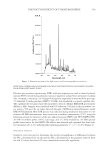
PHOTOCYTOTOXICITY OF TITANIUM DIOXIDE 537 White or lightly colored shades of inks were selected. The purchases were made between January 2006 and May 2010. All inks were explicitly labeled by the vendors for use as permanent makeup. GRAVIMETRIC DETERMINATION OF PERMANENT MAKEUP INKS’ PIGMENT CONTENT A 50-μl aliquot of permanent makeup ink was deposited onto a preweighed inorganic membrane fi lter (0.02-μm pore size, 10-mm diameter Whatman Inc., Clifton, NJ). The fi lter with deposited ink was weighed, and subsequently washed fi ve times under gentle vacuum using 50 μl of distilled water. The fi lter was then dried for three days in a ventilated oven operating at 35° ± 2°C. The fi lter was then weighed to obtain the weight of the dried pigment. The weight of the dried pigment and the initial weight of the permanent makeup ink were used to calculate the percentage of pigment in the permanent makeup ink. ISOLATION OF PIGMENTS FROM PERMANENT MAKEUP INKS One ml of permanent makeup ink was diluted with 3 ml of deionized water and centri- fuged at 85,000g (15°C) for one hour. (Optima L-90K ultracentrifuge, Beckman Coulter, Inc., Brea, CA). Sedimented pigments were washed twice by resuspension in 4 ml of deionized water and centrifugation as above. Pigments were then dried overnight under vacuum at 30°C. ELEMENTAL ANALYSIS OF PIGMENTS FROM PERMANENT MAKEUP INKS BY X-RAY FLUORESCENCE Each sample of pigment (approximately 200 mg) was mixed with paraffi n wax and pressed in a pellet die at 30 tons for fi ve minutes to form a standard pellet. X-ray fl uorescence mea- surements were made using a Bruker S4 wavelength dispersive X-ray fl uorescence spectrom- eter (Bruker AXS Inc., Madison, WI). The spectrometer sequentially searches for elements with atomic numbers from Na to U and adjusts the test conditions for each element to op- timize the detection sensitivity. A semiquantitative analysis was performed using the fl uo- rescence yield for each element and accounting for enhancements attributed to secondary excitation and absorption due to heavy elements. The semiquantitative analysis has a typical accuracy of 5%. The elements Al, Si, and Ti are reported as their most common oxides. DETERMINATION OF THE CRYSTALLINE PHASE OF TiO2 BY X-RAY DIFFRACTION Pigments isolated from permanent makeup inks were loaded onto a zero background holder and placed into a Phillips PW3020 diffractometer (Phillips Electronic Instru- ments, Inc., Mahwah, NJ) using Cu-Kα radiation at 40 KV and 30 mA. Scans were run over the range of 20° to 80° with a step size of 0.02° and a counting time of four hours. The crystalline phase of TiO2 was identifi ed using the powder diffraction fi le published by the International Centre for Diffraction Data (Newtown Square, PA). Once all phases were identifi ed, the amount of TiO2 in each was quantifi ed using a Rietveld refi nement for comparing the computed diffraction pattern with the observed diffraction pattern. CELL CULTURE Human skin fi broblasts (ATCC CRL-1634) were obtained from the American Type Culture Collection (Manassas, VA). Cells were cultured in Dulbecco’s modifi ed Eagle’s
Purchased for the exclusive use of nofirst nolast (unknown)
From: SCC Media Library & Resource Center (library.scconline.org)