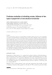
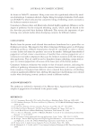
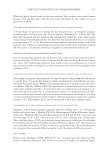
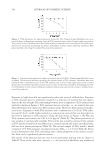
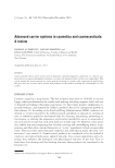
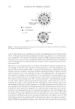
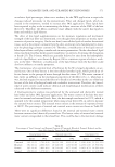
JOURNAL OF COSMETIC SCIENCE 538 medium, without phenol red, containing 10% fetal bovine serum, 50 μg/ml gentamicin, 4.5 mg/ml glucose, and 4 mM L-glutamine. All reagents used for cell culture were obtained from Invitrogen Corp., Carlsbad, CA. Cultures were incubated at 37°C in a humidifi ed atmosphere containing 5% CO2. IN VITRO ASSAY FOR CYTOTOXICITY AND PHOTOCYTOTOXICITY Fibroblasts were incubated for 18 hours with media containing either a permanent makeup ink or the pigment isolated from an ink. For treatment with a permanent makeup ink, a stock solution of the ink was prepared by dispersing the ink in deionized water us- ing a ten-second ultrasonic burst (Vibra-Cell VC250B sonicator, Sonics & Materials Inc., Danbury. CT). The stock solution was then heated for ten minutes at 100°C to minimize microbial contamination. Working solutions for treating fi broblasts were prepared by appropriately diluting stock solutions with media. Using the pigment content of each ink determined by gravimetric analysis, the level of treatment was expressed as the amount of pigment contained in the ink for each treatment and is given as μg of pigment per surface area of the fi broblast monolayer. For treatment with pigments, the pigments were dispersed in deionized water as described above and diluted in media to obtain working solutions used to treat fi broblasts. Following the 18-hour incubation in media containing a permanent makeup ink or pig- ment, fi broblasts were washed once with phosphate-buffered saline (PBS). Fibroblasts were then irradiated through freshly added PBS with 10 J/cm2 UVA radiation (320 nm– 400nm) combined with 45 J/cm2 visible light (400 nm–800 nm). Similar levels of UVA radiation and visible light would be received after exposure to the summer sun for 30 minutes (23). The source of UVA radiation and visible light was a 250-watt HITLite metal halide bulb (BLV Licht-und Vakuumteenik GmbH, Steinhöring, Germany) fi l- tered through glass. The emission spectrum of the light source was measured using an OL 754 UV-visible spectroradiometer (Optronic Laboratories Inc., Orlando, FL) and is shown in Figure 1. The spectral irradiance of the light source was found to be typically 6.3 × 10⁻³ W/cm2 UVA radiation and 2.8 × 10⁻² W/cm2 visible light. The emission of UVB radiation (280 nm–320 nm) from the light source was negligible (i.e., 1.1 × 10⁻⁷W/cm2). All irradiations were performed at 25° ± 3°C, and lasted approximately 25 minutes for simultaneous delivery of 10 J/cm2 UVA radiation and 45 J/cm2 visible light. To compen- sate for any inhomogeneity in the fi eld of illumination, uncovered samples were placed on a platform that rotated at 0.5 revolutions/min during irradiation. Sham-irradiated (i.e., dark control) samples were maintained at 25° ± 3°C in the dark. After irradiation, cells were removed from the dishes by trypsinization and plated into 60-mm Petri dishes (∼800 cells/dish). Four replicate dishes were plated for each treatment. The dishes were then incubated for 10–14 days to allow formation of cell colonies. Colonies were fi xed with methanol, stained with Giemsa stain, and counted. The average number of colonies observed after a treatment and the average number of colonies observed for cells receiving no treat- ment were used to calculate the percentage of cells surviving a treatment. Cytotoxicity was assessed using survival data for fi broblasts receiving treatment with an ink or a pigment alone (i.e., sham-irradiated samples), while photocytotoxicity was assessed from survival data for cells additionally exposed to light. A four-parameter logistic function (SigmaPlot 8, SPSS Inc., Chicago, IL) was used to fi t the data (i.e., % survival versus μg/cm2) and to determine the PD50 (dose of pigment which, in the presence of light, reduced survival by 50%).

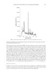
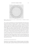
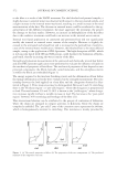
PHOTOCYTOTOXICITY OF TITANIUM DIOXIDE 539 DETECTION OF FREE RADICALS FORMED FOLLOWING PHOTOEXCITATION OF PIGMENTS ISOLATED FROM PERMANENT MAKEUP INKS Electron spin resonance spectroscopy (ESR) with spin trapping was used to detect hydroxyl radicals (HO•) formed during photoexcitation of pigments isolated from permanent makeup inks. A sample, containing 1 to 100 μg/ml of pigment suspended in water with the spin trap, 5,5-dimethyl N-oxide pyrroline (DMPO, 50 mM), was transferred to a quartz capillary tube. The capillary tube was placed into the microwave cavity of a Bruker EMX ESR spectrometer (Billerica, MA). Samples were irradiated with UV radiation (320 nm) in the microwave cav- ity, using a 500-watt Xe arc lamp directed through a McPherson monochromator, model DM200 (Chelmsford, MA). ESR spectra were collected during irradiation times from 1 to 25 minutes All ESR measurements were carried out at ambient temperature (27°C), using the following settings for detection of the spin adduct between DMPO and HO• (DMPO-OH): 20 mW microwave power, 100 G scan range, and 1 G fi eld modulation. An ESR spectral profi le characteristic for the DMPO-OH adduct was observed and contained four lines (rela- tive intensities of 1:2:2:1) with hyperfi ne splitting parameters aN = aH = 14.9 G (Figure 4). STATISTICAL ANALYSIS Student’s t-test was used to determine the statistical signifi cance of differences between the PD50 determined for an ink and the PD50 determined for the pigment isolated from the ink. P-values less than 0.05 were considered statistically signifi cant. Figure 1. Emission spectrum of the light source used for assessing photocytotoxicity.
Purchased for the exclusive use of nofirst nolast (unknown)
From: SCC Media Library & Resource Center (library.scconline.org)