JOURNAL OF COSMETIC SCIENCE 198 In 2008, Singh et al. (2) proposed the silver locus product Silv/gp100/Pmel17 as a new tool for the analysis of melanosome transfer in human melanocyte–keratinocyte co-cultures. In this study, they demonstrated, in a human in vitro model, that double im- munofl uorescent labeling of keratinocytes and melanosomes permits the defi nition of Pmel 17 as a tracker of transferred melanin and of the melanosome transfer. This work could have provided a good base for our own work, but double immnofl uorescent labeling and the quantifi cation of the obtained signals did not fi t well with our goal of a “quick and simple” method to quantify melanosome transfer. We therefore decided to combine the methodological approach of Singh et al. with other data concerning Pmel 17. Proteolytic ectodomain shedding of Pmel 17 has been described to lead to the release of a soluble part in the extracellular medium (3,4). We therefore reasoned that it should be possible to evaluate melanosome transfer by quantifying this “soluble” Pmel 17. Antibodies that indifferently recognize the melanosome-bound and/or the soluble form of Pmel 17 are now commercially available, and so we chose to develop an ELISA assay to quan- tify Pmel 17 in the extracellular medium of in vitro melanocyte–keratinocyte co-cultures. We decided to evaluate the effect of niacinamide, a well-known inhibitor of melanosome transfer (5,6), in order to validate our experimental design. This is the focus of the second part of this study. Finally, and in order to show that our methodological and technological approach can be easily and effi ciently used to achieve screenings and/or later phases of the development of new whit- ening compounds, we examined whether the whitening effect of an Alaria esculenta extract currently used in cosmetics could be due to the reduction of melanosome transfer. MATERIALS AND METHODS REAGENTS AND MATERIALS Human normal keratinocytes were obtained from a 45-year-old Caucasian donor. Human normal melanocytes were obtained from a 35-year-old Caucasian donor. Keratinocyte growth medium 2 (KGM 2) and melanocyte growth medium 2 PMA free (MGM 2 PMA free) were purchased from Promocell (Heidelberg, Germany). Penicillin and streptomy- cin came from Invitrogen (Carlsbad, CA). Melanoma gp100 protein (Pmel 17) was pur- chased from Abcam (Paris, France). BSA fraction V and Tween 20 came from Acros Organics (Geel, Belgium). Human melanocyte protein Pmel 17 (SILV) antibody came from US Biologicals (Swampscott, MA). The secondary antibody, i.e., a goat anti-chicken IgY (H&L) horseradish peroxidase conjugated antibody, was purchased from Antibodies- online (Aachen, Germany). The substrate reagent for the peroxidase came from R&D Systems (Minneapolis, MN). H2SO4 came from Merck (Darmstadt, Germany). Multiwell culture plates, Transwell® inserts, and EIA/RIA Stripwell plates were purchased from Corning Costar (Brumath, France). The Alaria esculenta extract came from BIOTECH- MARINE (Pontrieux, France). CELL CULTURES AND TREATMENTS Normal human adult melanocytes were seeded in multiwell culture plates and cultured in MGM PMA free in a humidifi ed incubator under a 5% CO2/95% air atmosphere

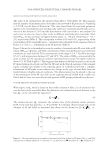
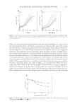
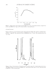
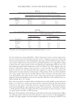
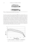
MELANOSOME TRANSFER EVALUATION 199 until they reached confl uence. Human normal adult keratinocytes were separately seeded in Transwell® inserts at 40000 cells per insert, 24 hours before the beginning of the experiments. For the study of the Pmel 17 production, Transwell® inserts containing the keratinocytes were transferred into the multiwell culture plates containing the melanocytes. The co- cultures were then incubated for a 72-hour period in the absence (control), or in the pres- ence of niacinamide at 5 mM or of an Alaria esculenta extract at 0.01% (v/v). At the end of the incubation period, the Pmel 17 was quantifi ed in culture media and the monolay- ers were rinsed with PBS before the quantifi cation of the cell lysates total protein content. Pmel 17 ELISA ASSAY Pmel 17 ELISA development. Multiwell plates specially designed to perform ELISA assays were incubated overnight at 4°C with 100 μl of serial dilutions (0, 1, 5, 10, 50, 100, 500, and 1000 ng/ml) of the standard peptide. In order to test different primary and secondary antibody dilutions, all the experimental conditions were performed in quadruplicate (n = 4). At the end of this incubation period, the wells were emptied and non-specifi c binding sites were saturated by the addition of 300 μl per well of a 1% BSA solution in PBS. After an incubation period of one hour at room temperature, the wells were emptied and fi lled with 100 μl of a 1% BSA solution in PBS containing the anti-human Pmel 17 an- tibody tested at two different concentrations (0.3 and 1 μg/ml). After an incubation pe- riod of two hours at room temperature, the wells were washed three times with PBS containing 0.1% of Tween 20. The wells were then fi lled with 100 μl of a 1% BSA solu- tion in PBS containing the secondary antibody (two different concentrations were tested: 1/1000 and 1/10000) coupled to a peroxidase, and were incubated at room temperature for a two-hour period. At the end of this incubation period, the wells were again washed three times with PBS containing 0.1% of Tween 20, and 100 μl of a solution containing a peroxidase substrate was added to each well. After 20 minutes, the peroxidase reaction was stopped by adding 50 μl of a 2N H2SO4 solution. The colorimetric signal was ana- lyzed (two wavelengths of reading: 450 and 550 nm) by using an appropriate spectropho- tometry plate reader (Victor V, Perkin Elmer, Waltham, MA). For calculations, the signal obtained at 550 nm was subtracted from those obtained at 450 nm. Pmel 17 ELISA assay in co-culture media. The multiwell plates specially designed to per- form ELISA assays were incubated overnight at 4°C with 100 μl of serial dilutions of the standard peptide or of the samples to be assayed. At the end of this incubation period, non-specifi c binding sites were saturated by the addition of 300 μl per well of a 1% BSA solution in PBS. After an incubation period of one hour at room temperature, the wells were emptied and fi lled with 100 μl of a 1% BSA solution in PBS containing the anti- human Pmel 17 antibody (antibody dilution: 1 μg/ml). After an incubation period of two hours at room temperature, the wells were washed three times with PBS containing 0.1% of Tween 20. The wells were then fi lled with 100 μl of a 1% BSA solution in PBS con- taining the secondary antibody (antibody dilution: 1/10000) coupled to a peroxidase, and were incubated at room temperature for a two-hour period. At the end of this incubation period, the wells were again washed three times with PBS containing 0.1% of Tween 20, and 100 μl of a solution containing a peroxidase substrate was added to each well. After
Purchased for the exclusive use of nofirst nolast (unknown)
From: SCC Media Library & Resource Center (library.scconline.org)