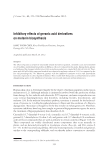
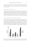
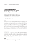
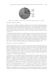
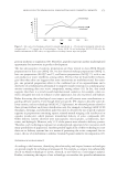
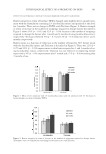
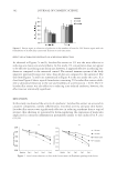
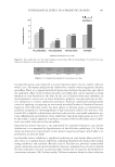
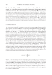
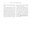
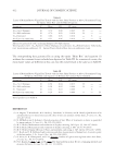
J. Cosmet. Sci., 63, 351–358 (November/December 2012) 351 Inhibitory effects of geranic acid derivatives on melanin biosynthesis SANG YOON CHOI, Korea Food Research Institute, Seongnam, Gyenoggi 463-746, Korea. Accepted for publication March 28, 2012. Synopsis The effects of geranic acid and its structurally related derivatives (geraniol, citronellic acid, and citronellol) on cell viability and melanin biosynthesis in Melan-a cells were evaluated in this study. Among them, geranic acid evidenced the strongest inhibitory activity on melanin production, coupled with low cell toxicity. Treat- ment with 500 μM of this compound resulted in a reduction in melanin content of 35.4% as compared to the live cell percentage (91.7%). Moreover, geranic acid also inhibited tyrosinase activity and intracellular tyrosinase expression in a dose-dependent manner. These results show that geranic acid may function as a skin depigmenting agent via the inhibition of tyrosinase activity and expression within melanocytes. INTRODUCTION Human skin color is determined largely by the degree of melanin pigment production in melanocytes (1,2). Although melanin is intimately involved with the protection of skin from damage by free radicals and ultraviolet (UV) exposure, melanin overproduction can cause serious hyperpigmentary skin disorders, including freckles, discoloration, and melasma (3,4). In the melanin biosynthesis pathway, tyrosinase is essential for the hydroxyl- ation of tyrosine to 3,4-dihydroxyphenylalanine (L-Dopa) and the oxidation of L -Dopa to dopaquinone. Tyrosinase is thought to be the key enzyme in melanogenesis (5). Therefore, tyrosinase inhibitors have long been sought as potential depigmentation agents for use in the treatment of hyperpigmentary skin disorders (6–8). Citronellol (3,7-dimethyl-6-octen-1-ol), citronellic acid (3,7-dimethyl-6-octenoic acid), geraniol (3,7-dimethylocta-2,6-dien-1-ol), and geranic acid (3,7-dimethyl-2,6-octadienoic acid) are fl avor compounds that are used as perfume in certain cosmetics (Figure 1) (9,10). Their compounds are widely distributed aroma components that occur naturally in plants, and are detected principally in Cymbopogon citratus, Rosa spp., and Vitis vinifera L (11–13). In the bioconversion pathway, the linear monoterpenoids, citronellol and geraniol, oxidize to the corresponding aldehyde, and can thereby induce conversion to citronellic Address all correspondence to S.Y. Choi at sychoi@kfri.re.kr.

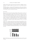
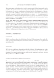
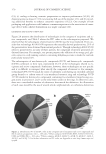
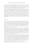
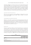
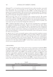
JOURNAL OF COSMETIC SCIENCE 352 acid and geranic acid by subsequent oxidation (14–16). Among these compounds, ge- ranic acid has recently been reported to exert a tyrosinase-inhibitory effect (11). However, no study has yet been conducted on its effects on melanocytes. The principal objectives of this study were to evaluate the inhibitory effects of these com- pounds on cell viability and melanin production in melanocytes, and also to assess their effects on the expression of melanin biosynthesis-associated enzymes, including tyrosinase. EXPERIMENTAL METHODS MATERIALS Citronellol, geraniol, citronellic acid, L -Dopa, and kojic acid were purchased from Sigma- Aldrich Co. (St. Louis, MO). Geranic acid was acquired from the Fluka Co. (Buchs, Switzerland). The Melan-a cell line was a gift from Dr. Bennett (St. George’s Hospital Medical School, London, UK). Fetal bovine serum (FBS), Roswell Park Memorial Insti- tute (RPMI) medium, and Penicillin–Streptomycin (PS) were purchased from Gibco BRL. (Grand Island, NY) CELL CULTURE PROCEDURES The Melan-a cells were cultured in RPMI 1640 medium under 10% FBS and 200-nm phorbol 12-myristate 13-acetate (PMA) conditions. In 100 ml culture dishes, 10 ml of medium was added and then seeded with 5 × 10⁵ cells. The cells were grown to confl u- ence after 3 to 4 days at 37°C in an atmosphere of 5% CO2, they were seeded with 105 cells/well in a 24-well plate, and then incubated for 24 h. Each well was replenished with 990 μl of medium daily, as well as treated with 10 μl of test sample [solvent (v/v): pro- pylene glycol/EtOH/H2O = 5/3/2] for 3 days the plate was then incubated for 1 day (17). CELL VIABILITY The percentage of viable cells was determined by staining the cell population using a simple crystal violet (CV) staining method to quantitate adherent cell number after treatment (18). After the removal of media from each well, the wells were washed with phosphate-buffered saline (PBS). Two hundred microliters of CV (0.1% CV, 10% EtOH, and the remaining volume as PBS) was then added. The plates were incubated at room temperature for 5 min Figure 1. Chemical structure of geranic acid and its derivatives.
Purchased for the exclusive use of nofirst nolast (unknown)
From: SCC Media Library & Resource Center (library.scconline.org)