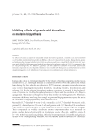
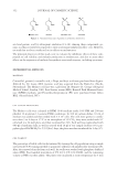
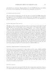
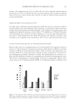
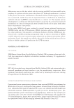
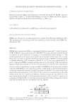
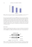
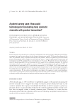
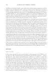
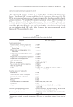
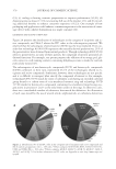
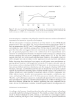
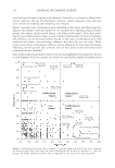
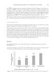
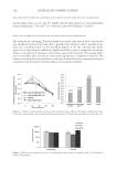
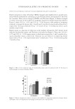
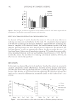
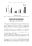
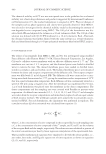
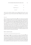
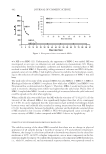
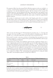
PROTECTION AGAINST UVB-INDUCED OXIDATIVE STRESS 361 HYDROGEN PEROXIDE DETERMINATION Hydrogen peroxide (H2O2) was measured as described previously (8). Briefl y, cells were treated with 10 μM 2′,7′-dichlorodihydrofluorescein diacetate (Molecular Probes, Eugene, OR) for 6 h and their fl uorescence measured (Ex485 nm /Em530 nm ). CELL VIABILITY Cell viability was evaluated by an MTS assay as described previously (9). MSRA AND METHIONINE SULFOXIDE PEPTIDE MSRA was obtained as an experimental test sample from Promega (Madison, WI). The pentapeptide containing metSO was synthesized and obtained from MMP Inc. (Plainfi eld, NJ). RESULTS NHEK were irradiated in Dulbecco’s phosphate-buffered saline, pH 7.4 with 50 mJ/cm2 UVB, the RNA isolated, and subjected to RT-PCR. The results from this analysis showed an increase in msrA expression in response to environmental trauma (Figure 1). The gapdh-normalized msrA levels (msrA/gapdh ratio) increased 22.6% from 0.53 to 0.65 (±0.07 S.E.). In order to establish whether increased amounts of MSRA could reduce ROS in NHEK following UVB irradiation, NHEK (2 × 10⁴) were pre-treated with 0.35 and 0.7 mg/ml of MSRA overnight and then exposed to 20 mJ/cm2 UVB. As shown in Figure 2, H2O2 decreased by 24.7% (±3.3% S.E.) and 36.1% (±0.5% S.E.), respectively. In a next series of experiments, an mp was added to NHEK for 24 h, harvested, and then analyzed by RT-PCR for msrA expression as before. Additionally, an identical pentapeptide containing methionine instead of metSO was tested in parallel. The results from these experiments demonstrated an 18.2% (±4.0% S.E.) increase in msrA expression in the cells treated with only 0.01 mg/ml mp, whereas a similar increase was not observed in the met-containing mp (Figure 3). Increases were observed at the 0.1 and 0.25 mg/ml con- centrations as well. Further, no cytotoxicity was measured for either pentapeptide as deter- mined by the MTS assay for cell viability (data not shown). Figure 1. Conventional RT-PCR analysis of UVB-irradiated NHEK showed an average increase in msrA expression when normalized to gapdh (0.53 vs. 0.65 ± 0.07 S.E.).

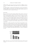
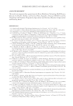
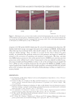
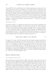
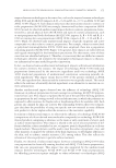
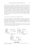
JOURNAL OF COSMETIC SCIENCE 362 To test the potential usefulness of boosting MSRA levels in skin, mp was then added to the media of skin models for 48 h, exposed to 100 mJ/cm2 UVB, and then incubated for an additional 24 h post-irradiation. The skin model samples were fi xed, subjected to histological analysis, and stained with hematoxylin and eosin. In Figure 4, light micro- scopic images of these skin models show an increase in sunburn cells in UVB-exposed skin model samples. Sunburn cells are characterized by increased pyknotic nuclei and eosinophilic staining (10). In contrast, the presence of sunburn cells in skin model samples pre-treated with the mp prior to UVB exposure was signifi cantly diminished by 31.1% (±10.0% S.E.). DISCUSSION Human skin has developed many strategies to counteract the effects of environmental exposures and their consequent trauma. In this report, we describe the presence of msrA in NHEK and its induction by UVB. Our data indicate a rapid and protective Figure 2. H2O2 levels decreased in the presence of 0.35 and 0.70 mg/ml MSRA by 24.7% (±3.3% S.E.) and 36.1% (±0.5% S.E.), respectively (∗ = Student’s t-test assuming unequal variances, p 0.05, n = 3). Figure 3. Conventional RT-PCR analysis of NHEK pre-treated with mp. msrA expression increased by 18.2% (±4.0% S.E., n = 4) when treated with 0.01 mg/ml mp and normalized to β-microglobulin. Higher levels (0.1 and 0.25 mg/ml) of mp also induced increases in msrA, whereas the unoxidized form had no effect.
Purchased for the exclusive use of nofirst nolast (unknown)
From: SCC Media Library & Resource Center (library.scconline.org)