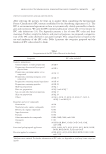
NICOTINAMIDE MICROEMULSION-BASED GELS 399 Acetonitrile, triethylamine, and methanol, which were used in high performance liquid chromatography (HPLC) assay, were supplied by Lab-Scan Analytical Science (Bangkok, Thailand). Distilled water was used throughout the experiments. All chemicals were of pharmaceutical grade and used without further modifi cation. A commercial cream (CC) containing 3% w/w of nicotinamide was purchased from a local supermarket in Songkhla Province, Thailand. Cellulose acetate membrane (Spectra/Por®3 dialysis membrane, MWCO 3500 Dalton) was obtained from Spectrum Laboratories Inc. (Los Angeles, CA). Polyamide membrane fi lter was obtained from Sartorius AG (Göttingen, Germany). PREPARATION OF MICROEMULSION-BASED GELS ME-2 from our previous study (16), referred to as ME here, was further used for converting to three nicotinamide MBGs designated as MBG-1, MBG-2, and MBG-3 via the formu- lations as shown in Table I. CHARACTERIZATION OF MICROEMULSION-BASED GELS Appearance of the samples was optically observed. Viscosity and class of fl ow of the samples were measured at fi ve different speeds using a Brookfi eld DV-III Ultra rheometer (Brookfi eld Engineering Laboratories Inc., Middleboro, MA) fi tted with an LV spindle. Brookfi eld Rheocalc operating software (version 3.1-1) was used to control the rheometer. The determinations were performed at 32°C, equal to the temperature of human skin. The measurements were performed in triplicate. STABILITY STUDY The physical and chemical stabilities of the prepared formulations were investigated by storing the samples at three temperatures—4°C, ambient temperature (approximately 30°C), and 60°C—for 2 months. The 4°C represented the temperature of a refrigerator, the ambient temperature represented the normal usage as well as storage conditions, and 60°C mimicked the temperature on transportation during summer in Thailand. Since the temperature during summer in Thailand is fl uctuated high, it is interesting to investigate the changes in the appearance of MBGs when stored at high temperature in this study. Table I Formulations of the Investigated MBGs Component % w/w MBG-1 MBG-2 MBG-3 ME 95 95 95 Colloidal silica 5 — — Carbomer (0.5% w/w solution in water, adjusted the pH to 6 with TEA) — 5 3 PEG-40 hydrogenated castor oil — — 2

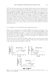
JOURNAL OF COSMETIC SCIENCE 400 The chemical stability at 60°C was not anticipated since in the guideline for accelerated stability test of new drug substances and products suggested by International Conference on Harmonization (17), the studied temperature is assigned at 40°C. Physical changes of the samples such as phase separation and clarity were optically observed. Only MBG-1 was selected to investigate for the chemical stability study by determining the amount of non-degraded active compound using HPLC. The 100 μl of the formulation was dis- solved with IPA and adjusted the volume in a 10-ml volumetric fl ask. The 100 μl of this solution was diluted with 80:20 IPA/methanol in a 10-ml volumetric fl ask. Afterward, the obtained solution was further diluted with the same solvent in a 10-ml volumetric fl ask and then fi ltered through a 0.45-μm polyamide membrane fi lter before HPLC analysis. IN VITRO RELEASE STUDY The release of nicotinamide from MBG-1, ME, and CC was investigated using modifi ed Franz diffusion cells (Hanson Model 57-6 M, Hanson Research Corporation, Los Angeles, CA) and a cellulose acetate membrane with an effective diffusion area of 1.77 cm2. The membrane was cut into 3 × 3 cm pieces, and the obtained pieces were boiled in distilled water to remove the wax. The cleaned membrane pieces were soaked in distilled water, stored in a cool place, and used within a week. The hydrated membrane pieces were mounted between the donor and receptor compartments of the diffusion cells. The receptor compart- ment was fi lled with 11 ml of degassed IPB. The diffusion cells were connected to a circu- lating water bath thermostated at 37°C, giving the membrane surface temperature of 32°C that was equal to human skin temperature. Each diffusion cell was continuously stirred at a speed of 200 rpm using a magnetic bar. After the membrane was equilibrated for 30 min, 1 g of each formulation was placed onto the membrane in the donor compartment. The donor compartment and the sampling arm were covered with Parafi lm to prevent water evaporation. At defi ned time intervals (0.5, 1, 2, 4, 8, 10, 12, and 25 h), 0.5 ml of samples were taken from the receptor compartment and immediately replaced with an equal volume of fresh receptor medium. The withdrawn samples were analyzed for nicotinamide concen- tration by HPLC. For each formulation, the experiment was performed in triplicate. The cumulative release (Q) of nicotinamide was calculated from equation (1). ¦V 1 0 t r t s i i Q V C C (1) where Ct is the concentration of active compound in the receptor fl uid at each sampling time t, Ci is the concentration of active compound of the ith sample, and Vr and Vs are the volumes of the receptor fl uid and the sample, respectively. The release rate was calculated as percent of the initial concentration per hour by linear regression interpolation of the experimental data. Three possible mathematical equations were employed to fi t with the release profi les, i.e., zero order, fi rst order, and Higuchi square root of time equations as shown in equations (2), (3), and (4), respectively.
Purchased for the exclusive use of nofirst nolast (unknown)
From: SCC Media Library & Resource Center (library.scconline.org)