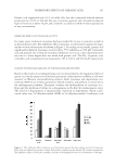
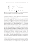
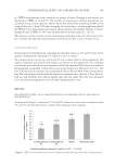
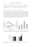
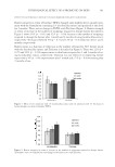
PHYSIOLOGICAL EFFECT OF A PROBIOTIC ON SKIN 387 Oil-in-water (o/w) formulations were prepared at 1% and 5% concentrations of the pro- biotic for the studies. Triclosan (BASF, NC), a known antibacterial, at 0.1% concentra- tion prepared in a similar formulation base, was used as a control for some parts of the study. In addition, a similar formulation base with 1% salicylic acid (Rhodia Inc., NJ) was used as an internal control for the acne study presented in this paper. All clinical studies were conducted following good clinical practice standard (ICH Topic E 6-R1 July 2002 CPMP/ICH/135/95). The subjects recruited in these studies were in normal health with no evidence of acute or chronic disease other than acne. Written in- formed consent was obtained from each volunteer before entering into the study. The subjects were not on any antibiotic, antihistamines, retinoid, anti-infl ammatories or steroid therapy, benzoyl peroxide, and/or salicylic acid treatment for at least 2 weeks prior to commencement of this study. The subjects were not under the care of a der- matologist and were not on any acne treatment for at least 1 month before the study started. Pregnant or lactating females were excluded, also subjects exhibiting current sunburn, rashes, scratches, burn marks, etc., which might interfere with the evaluation of test results. SKIN SENSITIVITY The anti-infl ammatory properties of the test materials were tested by observing the re- duction of onset and intensity of erythema induced by an irritant, Balsam of Peru. The test site was on the volar forearms of subjects with a history of sensitivity to Balsam of Peru (15). “Balsam of Peru” (8% w/w in petrolatum), an irritant that contains approxi- mately 0.9% cinnamic aldehyde (16,17), was applied at a dose of approximately 4 mg/cm2. Erythema was measured with a Minolta Chromameter (Konica Minolta, Ramses, NJ). Part I: Reduction of onset of skin redness. Ten subjects with a history of skin sensitivity to Balsam of Peru were chosen for the study. The test compounds were applied on the volar forearms of the subjects. The material was allowed to absorb for 30 min and then Balsam of Peru, the irritant, was applied on the test sites. When erythema appeared, even on one site, the arms were washed with wet towels and skin redness was measured with the Chromameter. Red (a∗ values) subtracted by the baseline skin redness determined an “increase in redness (Δa∗) due to irritation.” A comparison of Δa∗ with the positive and negative controls exhibited the potential of the test materials for reducing the onset of skin irritation. The positive control was a∗ values of skin treated with Balsam of Peru alone. Part II: Reduction of intensity of skin redness. A total of ten subjects participated in the study. Using a pen, 1.5 in.2 areas were marked on each volar forearm of the subjects correspond- ing to the test materials and the positive and negative controls. Baseline color measure- ments were obtained from all the sites using a Minolta Chromameter. Balsam of Peru (8% pet) was applied on all the sites at the rate of approximately 4 mg/cm2 in a 1.5-cm diameter circle. When redness appeared approximately evenly on all the sites, the irritant was wiped off with a wet towel and then washed with warm water. The degree of redness was measured with the Chromameter on all the sites as the baseline redness. The test materials were applied on their respective sites at the rate of 2 mg/cm2 and color measurements were obtained after 15 min, 30 min, 1 h, 1.5 h, and 2 h.

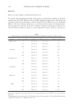
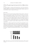
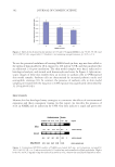
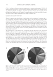
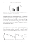
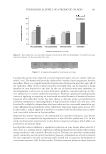
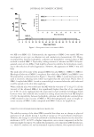
JOURNAL OF COSMETIC SCIENCE 388 Increase in color was determined by subtracting the baseline skin color values of all the sites and color values were plotted against time. Area under the curve was obtained for all the sites. SKIN BACTERIAL COUNT The test materials used in this section were an o/w base cream containing lactobacillus extract (L. plantarum) at 1% and a similar formulation with Triclosan (an antibacterial) at 0.1% as a control. A total of 29 females between the age of 25 and 55 participated in the study. The panel was divided into three groups of 9–10 each. The subjects were provided with the test material to use twice a day for 2 months on the full face. They were instructed not to use any other moisturizers or treatment products however, they could continue to use the cleansers and makeup that they normally use as long as they did not change products during the course of the study. On the day of the study, the subjects reported to the lab with a clean face and forearms, with no creams, lotions, makeup, etc. Skin microfl ora measurements described below were obtained at baseline, 1 month, and 2 months. Skin microfl ora. The subjects (n = 29) reported to the laboratory and washed their face with a (non-antibacterial) mild liquid soap. Normally, after washing with soap and water, the bacterial count of skin drops to almost zero with gradual increase in microfl ora over time. The normal microfl ora was allowed to appear on the skin for the next 3 h. During this pe- riod, the subjects were advised to keep their hair away from the forehead and refrain from touching the face or wash it or apply anything on it. At the end of this 3-h time point, saline washings were obtained from the forehead of the face for microbiological analysis. Dulbeccos phosphate-buffered saline (PBS) washings of the forehead area were obtained using a sterile glass cylinder and a sterile rubber policeman. One milliliter of the saline was poured in the cup and then the skin was scrubbed with rubber policeman (10 strokes) and washed and then the saline was aspirated and collected in 9 ml of PBS. The samples were analyzed for aerobic and anaerobic bacterial count. The data were averaged to deter- mine the total microfl ora. The samples were analyzed for microfl ora as per the U.S. Pharmacopeia Chapter 61 (18) where 1:10 dilutions of the samples were prepared in PBS and plated on tryptic soy agar. After 48 h of incubation at 37°C, the plates were examined and the recovered organisms were quantifi ed. All recovered organisms were then streaked for single colony isolation, gram stained, and examined under the microscope. The organisms were identifi ed using the Becton–Dickinson BBL Crystal™ Identifi cation System. BARRIER FUNCTIONS The test site was the jawline of the face of the subjects. Basal skin barrier was determined by measuring trans epidermal water loss (TEWL), using a Servomed Evaporimeter (Ser- voMed AB, Stockholm, Sweden). To determine barrier integrity, a tape (Tuck tape) was used to cover the test area and after a fi rm stroke in both directions the tape was peeled off (19). A total of three strippings were obtained. TEWL was recorded again. Strippings followed
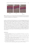
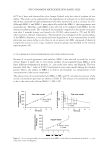
Purchased for the exclusive use of nofirst nolast (unknown)
From: SCC Media Library & Resource Center (library.scconline.org)