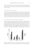
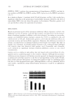
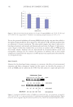
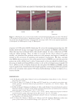
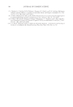
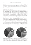
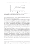
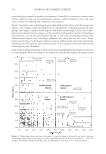
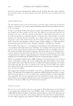
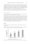
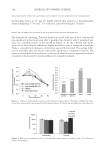
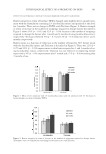
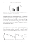
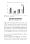
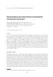
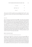
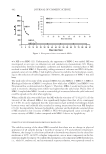
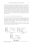
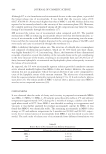
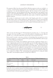
PHYSIOLOGICAL EFFECT OF A PROBIOTIC ON SKIN 393 Lactobacillus extract also impacted a second important aspect of acne, namely infection with P. acnes. The human skin provides a habitat for a variety of microorganisms: the skin microfl ora. There is a complex network of interactions between the microbes and cells of the epidermis. Most of the resident microbes on healthy skin can be regarded as being harmless or even benefi cial to the skin. In the case of diseases with some imbalance in microorganisms, such as acne or atopic dermatitis, probiotic concepts represent an effec- tive alternative to strictly antibacterial products. Probiotic approaches predominantly consist of applying or ingesting an inactivated microbial biomass of benefi cial bacteria. Ingestion of lactobacillus extract has been shown to alleviate atopic eczema/dermatitis syndrome symptoms in immunoglobulin E (IgE)-sensitized infants (25) and mice (26). Lactobacillus acidophilus cultures have also been advocated as a successful remedy for sys- temic infl ammation and oxidative stress, which have important implications in acne (27). In this study, a topical approach to probiotic treatment with lactobacillus extract exhib- ited a successful reduction in skin microfl ora. Impaired skin barrier function is also responsible for comedone formation, since barrier dysfunction is accompanied by hyperkeratosis of the follicular epithelium (11). In this study, we observed an improvement in skin barrier integrity and repair, which adds to its potential as an antiacne agent. Lactobacillus extract exhibited a signifi cant reduction in acne lesions when used for 4 days. Acne is a common disease, which has confounded hundreds of remedies that include strong antibiotics and retinoids. Biocides such as Triclosan are powerful antimicrobial agents commonly used in topical treatments, however, widespread use of such a material has been reported to become a potential public health risk in regard to development of concomitant resistance (28). In these studies, Triclosan treatment showed a robust reduction Figure 8. Area under the curve size and erythema of acne lesion. The data from Figure 7(a) and (b) are sum- marized in Figure 8 as the area under the curve. Figure 9. A sample photograph of acne lesion over time.

JOURNAL OF COSMETIC SCIENCE 394 in bacterial count after 1-month use however, after 2 months of use, there was a marked increase in the microfl ora, suggesting the possibility of resistance. Although bacterial resistance to antibiotics is rampant, much is not known about the effect of long-term use of lactobacillus extract nevertheless, it promises to be a valuable agent in the reduction of the intensity of acne lesions. REFERENCES (1) E. Metchnikoff, The Prolongation of Life, Optimistic Studies (Putnam’s Sons, New York, 1908), pp. 161– 183. (2) D. M. Lilly and R. H. Stillwell, Probiotics: growth-promoting factors produced by microorganisms, Science, 147, 747–748 (1965). (3) P. Marteau, E. Cuillerier, S. Meance, M. F. Gerhardt, A. Myara, M. Bouvier, C. Bouley, F. Tondu, G. Bommelaer, and J. C. Grimaud, Bifi dobacterium animalis strain DN-173 010 shortens the colonic transit time in healthy women: a double-blind, randomized, controlled study, Aliment Pharmacol Ther., 16, 587–593 (2002). (4) B. S. Gan, J. Kim, G. Reid, P. Cadieux, and J. C. Howard, Lactobacillus fermentum RC-14 inhibits Staphylococcus aureus infection of surgical implants in rats, J Infect Dis., 185(9), 1369–1372 (2002). (5) B. Karska-Wysocki, M. Bazo, and W. Smoragiewicz, Antibacterial activity of Lactobacillus acidophilus and Lactobacillus casei against methicillin-resistant Staphylococcus aureus (MRSA), Microbiol Res., 165(8), 674–686 (2010). (6) R. S. Ali, A. Falconer, M. Ikram, C. E. Bissett, R. Cerio, and A. G. Quinn, Expression of the peptide antibiotics human beta defensin-1 and human beta defensin-2 in normal human skin, J Invest Dermatol., 117(1), 106–111 (2001). (7) M. Frye, J. Bargon, and R. Gropp, Expression of human beta-defensin-1 promotes differentiation of keratinocytes, J Mol Med., 79(5–6), 275–282 (2001). (8) T. Tomita and T. Nagase, Defensins as a mechanism of host defense and innate immunity, Nippon Ronen Igakkai Zasshi, 38(4), 440–443 (2001). (9) Y. Sang and F. Blecha, Antimicrobial peptides and bacteriocins: alternatives to traditional antibiotics, Anim Health Res Rev., 9(2), 227–235 (2008). (10) H. Baba, A. Masuyama, C. Yoshimura, Y. Aoyama, T. Takano, and K. Ohki, Oral intake of Lactobacillus helveticus-fermented milk whey decreased transepidermal water loss and prevented the onset of sodium dodecylsulfate-induced dermatitis in mice, Biosci Biotechnol Biochem., 74(1), 18–23 (2010). (11) A. Yamamoto, K. Takenouchi, and M. Ito, Impaired water barrier function in acne vulgaris, Arch Der- matol Res., 287(2), 214–8 (1995). (12) N. Muizzuddin, “Acne: Causes, Treatment and Myths,” in Dermatology Research Focus on Acne Melanoma and Psoriasis, D. E. Roth. Ed. (Nova Science Publishers, New York, 2009), pp. 97–106. (13) P. Lyte, R. Sur, A. Nigam, and M. D. Southall, Heat-killed Propionibacterium acnes is capable of inducing infl ammatory responses in skin, Exp Dermatol., 18(12), 1070–1072 (2009). (14) M. Sullivan, S. Schnittger, T. Mammone, and E. Goyarts, Skin treatment method with lactobacillus extract, U.S. Patent 7,510,734,B2 (2009). (15) N. Muizzuddin, K. D. Marenus, and D. H. Maes, Factors defi ning sensitive skin and its treatment, Am J Contact Dermat., 9(3), 170–175 (1998). (16) C. Matthies, A. Dooms-Goossens, J. M. Lachapelle, A. Lahti, T. Menne, I. R. White, and J. Wilkinson, Patch testing with fractionated balsam of Peru, Contact Dermatitis, 19, 384–385 (1988). (17) B. M. Hausen, T. Simatupang, G. Bruhn, P. Evers, and W. A. Koenig, Identifi cation of new allergen constituents and proof of evidence for coniferyl benzoate in Balsam of Peru, Am J Contact Dermat., 6(4), 199–208 (1995). (18) The United States Pharmacopeial Convention, Microbial Limit Tests in United States Pharmacopeia, 27th revision/National Formulary, 22th Ed. (The United States Pharmacopeial Convention, Inc., Rockville, MD, 2003). (19) P. M. Elias, Epidermal lipids, barrier function, and desquamation, J Invest Dermatol., 80 (Suppl), 44s– 49s (1993). (20) G. Grubauer, P. M. Elias, and K. R. Feingold, Transepidermal water loss: the signal for recovery of bar- rier structure and function, J Lipid Res., 30(3), 323–33 (1989).
Purchased for the exclusive use of nofirst nolast (unknown)
From: SCC Media Library & Resource Center (library.scconline.org)