JOURNAL OF COSMETIC SCIENCE 80 Fibroblasts are cells that grow in the dermal layer of the skin and are responsible for expression of new collagen and elastin. Dermal papillae cells also grow in the dermis of the skin and are the cells responsible for expression of hair fi bers. Dermal papillae cells from balding and non-balding individuals have recently been grown ex vivo (5). Dermal papillae cells from balding individuals exhibit signature protein markers in- dicating they are experiencing premature senescence. They have been found to express high levels of senescence-associated β-galactosidase (SA-β-Gal) and ataxia telangiecta- sia mutated (ATM) proteins, both being accepted markers for cellular senescence (5). Ultraviolet radiation (UVR)–stressed fi broblasts from aged skin also express high lev- els of these two proteins suggesting senescence plays a strategic role in extrinsic skin aging as well (6). For fi broblasts from aged skin, it has been determined that they express high levels of both ATM protein and SA-β-Gal indicating these cells are ap- proaching or have reached cellular senescence (7). Phenotypically, aging includes effects from the subcellular to macroscopic level. Such signs of aging may be induced or caused by intrinsic factors, e.g., chronological ag- ing, or extrinsic factors, e.g., environmental damage, sunlight, UV, smoke, ozone, pollutants, stress, etc. Visible signs of skin aging include an increase of fi ne lines, wrinkles, large pores, and surface roughness. Recently, it was reported that in mice genetically modifi ed to show accelerated senescence that selective removal of senes- cent cells demonstrated a signifi cant improvement in the aging phenotype of the mice compared against controls in which the senescent cells were not removed (8). The study demonstrates that even though senescent cells make up only a small per- centage of the total cells within the mice, the infl uence of these senescent cells on aging is profound. Biochemical markers for replicative senescence have been identifi ed and include the genes p53 (TP53), Sirtuin1 (SIRT1), ataxia telangiectasia mutated (ATM) and the re- lated ataxia telangiectasia related protein (ATR) (9). In addition, Rad23 (RAD23), p21 (TP21), and p16 (TP16) have also been identifi ed as important markers for cel- lular senescence. ATM protein plays a strategic role in DNA checkpoint response functions linked in part to ATM-directed phosphorylation/activation of p53 and a host of other cellular DNA-damage response proteins (10–12). ATM protein also plays a central role in signaling the presence of DNA double-strand breaks. Loss of ATM protein function is characterized by accelerated telomere loss, genomic instability, progressive neurological degeneration, and premature aging. ATM protein defi ciency and telomere dysfunction likely act together to impair cellular and whole-organism viability. High ATM protein expression is associated with cells in senescence. Changes in expression of ATM protein can also be used as a biomarker to identify senescent cells. Cellular senescence can be observed via various methods. Cellular senescence leads to an increase in SA-β-Gal activity, which can be used as a biomarker to identify senes- cent cells (13). SA-β-Gal is expressed by cells in either intrinsic or stress-induced cellular senescence. Senescent cells can also be noted by changes in the morphology of the cells. It has been reported that extracts from yeast fermentation, in particular, Saccharomyces cerevisiae, have demonstrated wound healing properties (14–19). These physiological effects, which have been variously attributed to increased cellular oxygen consumption

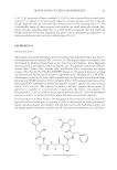
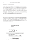
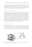
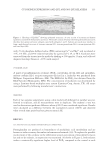
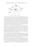
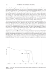
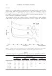
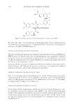
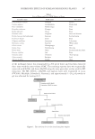
MODULATION OF CELLULAR SENESCENCE 81 (14,17,19), improved collagen synthesis (15,18,19), and increased blood vessel devel- opment (17), appear to be principally related to unique proteins and low molecular weight peptides that are enzymatically manufactured in the growing yeast (17,18). Undoubtedly, many of these proteins and peptides are small molecular weight frag- ments of larger signaling molecules. It is now widely recognized that low molecular weight proteins and nuclear fragments can play a role in upregulating important cel- lular growth factors that can lead to skin healing (20–23). EXPERIMENTAL PEPTIDE ISOLATION The peptide used in the following studies was isolated and identifi ed from a low molecu- lar weight fraction of a ferment of S. cerevisiae (24). The peptide amino acid sequence was determined by Erdman Degradation at the University of California, Davis Molecular Structure Facility and indicated that the peptide was a hexapeptide comprised of Phenyl- alanine (Phe), Valine (Val), Alanine (Ala) and Proline (Pro), comprising the unique sequence Phe-Val-Ala-Pro-Phe-Pro (FVAPFP) (Figure 1). The peptide has been assigned the International Nomenclature Cosmetic Ingredient name, Hexapeptide-11. BLAST2® (Washington University, St Louis, MO) available with the National Center Biotechnology Information (NCBI) Sequence Viewer software (http://www.yeastgenomeorg/cgi-bin/ blast-sgd.pl) was employed to match this peptide sequence against the entire protein dataset for S. cerevisiae. The sequence of amino acids that comprise Hexapeptide-11 appears in a number of yeast proteins in particular, the amino acid sequence for Hexapeptide-11 can be found in stress-related proteins (hsp70), and transmembrane proteins as well as a number of proteins whose functions are presently unknown. For the purposes of these studies, the hexapeptide was synthesized using solid state peptide synthesis techniques to a purity of 95% as determined by high-performance liquid chromatography (not shown). The peptide used in the following studies was the highly purifi ed synthetic peptide and all concentrations shown are on a dry basis. Figure 1. Structure of Hexapeptide-11.
Purchased for the exclusive use of nofirst nolast (unknown)
From: SCC Media Library & Resource Center (library.scconline.org)