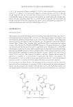
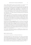
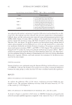
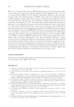
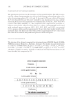
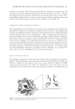
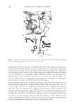
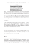
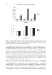
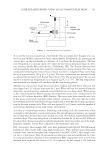
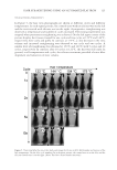
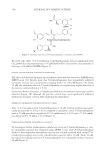
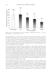
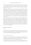
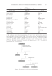
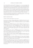
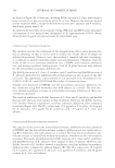
JOURNAL OF COSMETIC SCIENCE 102 PURIFICATION OF THE TYROSINASE INHIBITOR The purifi cation was based on the tyrosinase activity-guided method. The EA was sepa- rated by column chromatography over silica gel with mixtures of petroleum ether–acetic ether of increasing polarity (9:1–4:6), and 14 fractions (A–N) were collected. Fractions A–F were discarded because they were insoluble in our enzyme activity assay, and the remaining 8 fractions were separated over a Sephadex LH-20 (GE Inc., New York, NY) column with CHCl3–MeOH (1:1). The compounds with a high level of tyrosinase in- hibition were further refi ned by C18 (GE Inc.) column chromatography and eluted with methanol aqueous solutions (20%, 40%, 60%, 80%, and 100%, successively) (v/v) to prepare a white powder (approximately 30 mg), which was washed with the 40% methanol eluent. The fi nal yield of the white powder is approximately 0.01%. The overall fl ow chart of the isolation and purifi cation process is shown in Figure 1. STRUCTURE DETERMINATION The structure of the obtained compound was determined using AVANCE Digital 400 MHz NMR Spectrometer (Bruker Inc., Karlsruhe, Germany). The detailed structural information is as follows: 1 H-NMR (CDCl3,δ, ppm.) 7.56 (1H, d, J = 4 Hz), 7.54 (1H, d, J = 4 Hz), 6.83 (1H, t, J = 8 Hz), 3.90 (3H, s) 13 C-NMR (CDCl3, δ, ppm.) 170.21(s), 152.85(s), 148.85(s), 125.48(d), 123.29(s), 116.04(d), 114.08(d), and 56.63(q). Previous reports (17,18) have identifi ed this compound as VA. Figure 1. Flow chart of isolation of tyrosinase inhibitors from red globe amaranth.

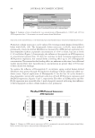
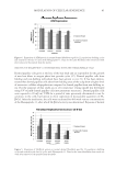
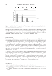
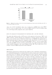
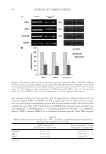
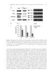
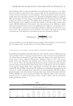
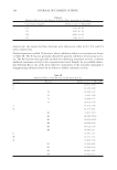
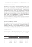
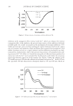
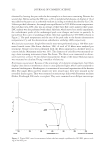
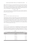
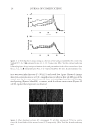
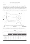
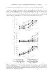
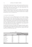
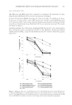
INHIBITORY MECHANISM OF RED GLOBE AMARANTH ON TYROSINASE 103 CIRCULAR DICHROISM MEASUREMENTS Circular dichroism (CD) spectroscopy can be used to estimate the overall conformation of protein molecules. The CD measurements of tyrosinase in the presence and absence of VA were obtained in the range of 190–250 nm using a 2-mm quartz cuvette at a scan speed of 60 nm/min, with the results from three scans averaged for each CD spectrum. Each 1.0 ml mixture contained 0.9 ml tyrosinase (0.1 mg, dissolved in water) and 0.1 ml VA (dissolved in a 10% methanol aqueous solution). The VA/tyrosinase molar ratio was varied (0, 1:1, and 4:1), and the CD spectra were recorded using MOS-450 spectrometer (Bio- Logics Inc., Grenoble, France) at 25°C (19,2 0). ULTRAVIOLET SPECTROSCOPIC MEASUREMENTS Ultraviolet (UV)/visible spectroscopy was used to evaluate whether VA could chelate copper ion of tyrosinase. Spectra at 240–400 nm were measured using the UV-2102 spectrophotometer (Unico Inc., Shanghai, China). The mixture contained 1.9 ml of the samples (10 μg/ml, dissolved in 0.2% methanol) and 0.1 ml of 50 mM phosphate buffer (pH 6.8) with mushroom tyrosinase (100 units/ml). Scans of 1.0 mM CuSO4 were obtained for comparison (21,22). MODEL BUILDING AND MOLECULAR DOCKING The crystal structure of tyrosinase from Bacillus megaterium (TyrBm PDB ID: 3NQ1) (23) was chosen as the protein model in this study. The dicopper and ligands were re- moved from 3NQ1 before the docking computation was performed. The docking algo- rithm was based on the ROSETTALIGAND software (http://www.rosettacommons.org) as previously described (24,25). For each receptor–ligand pair, 500 docking results were generated from the docking calculation. Then, top 10 structures were selected based on the total ROSETTA energy, and they were ranked by the receptor–ligand interaction energy and ligand conformational variation parameters. The fi gures were produced using the PyMOL molecular graphics system (http://www.pymol.org). RESULTS AND DISCUSSION EFFECT OF FRACTIONS FROM RED GLOBE AMARANTH ON TYROSINASE ACTIVITY Table II shows the inhibitory effect of the crude extracts from red globe amaranth fl ower on tyrosinase. Table II shows that the crude extract had a concentration-dependent in- hibitory effect on tyrosinase activity, with an IC50 value of 3.32 mg/ml. These results suggest that the crude extract of red globe amaranth fl ower has potential inhibitory ef- fects on tyrosinase. The inhibitory effects on tyrosinase by additional EA extract fractions are presented in Table III, which shows that the eight fractions (G–N) inhibited tyrosi- nase activity to varying degrees. Overall, the fractions I, K, and N could inhibit tyrosi- nase activity considerably with inhibition rates of 51.05%, 54.37%, and 70.89%,
Purchased for the exclusive use of nofirst nolast (unknown)
From: SCC Media Library & Resource Center (library.scconline.org)