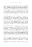
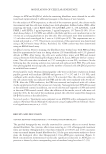
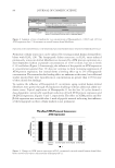
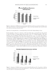
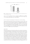
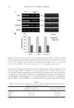
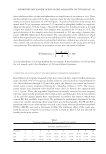
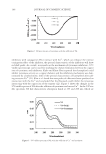
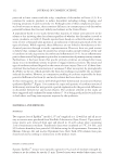
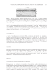
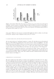
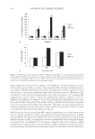
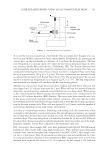
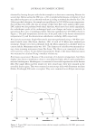
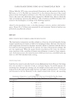
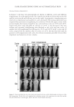
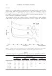
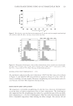
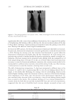
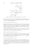
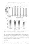
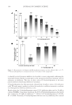
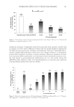
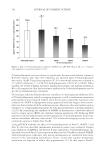
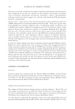
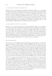
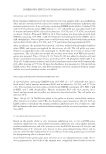
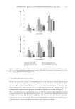
JOURNAL OF COSMETIC SCIENCE 106 inhibitors with conjugative effect interact with Cu2+, which can enhance the interior conjugation effect of the inhibitor, the spectral characteristics of the inhibitors will show red shift peaks. As a result, in research on the mechanism of tyrosinase inhibitors , UV/ visible spectroscopy can be used for studying the chelate formation between the copper ions of tyrosinase and inhibitors. Kubo and Kinst-Hori reported that kaempferol could inhibit tyrosinase activity as a copper chelator, and the inhibitory mechanism was dem- onstrated by a bathochromic shift of the spectral characteristics of kaempferol after add- ing excessive Cu2+ (21). Kim et al. found that new peaks of a fl avonoid were produced on interaction with the Cu2+ and concluded that the fl avonoid could inhibit the tyrosinase interacting with the copper ion of the enzyme (22). Figure 3 shows the changes in the UV/visible spectra of VA after the addition of tyrosinase and excess Cu2+. In the UV/vis- ible spectrum, VA had characteristic absorption bands at 255 and 288 nm, which are Figure 2. CD spectroscopy of tyrosinase with the addition of VA. Figure 3. UV/visible spectrum of VA with the addition of Cu2+ and tyrosinase.

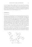
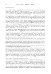
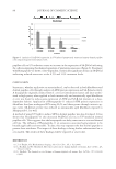
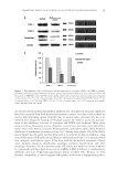
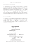
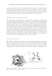
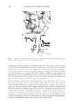
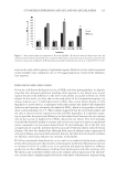
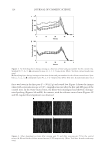
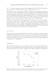
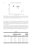
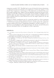
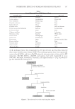
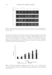
INHIBITORY MECHANISM OF RED GLOBE AMARANTH ON TYROSINASE 107 assumed to be band K (210–250 nm) and band B (260–300 nm) of a benzene ring. The maximum absorptions of VA remained the same after the addition of excess Cu2+, whereas the absorption maxima shifted to 249 and 285 nm after the addition of tyrosinase. These results illustrate that the VA can interact with tyrosinase however, chelation interaction between VA and the dicopper copper ion of the enzyme is not included (30). STEREOSCOPIC STRUCTURAL HOMOLOGY ANALYSIS The conformations of VA docked onto tyrosinase were investigated to determine the mechanism of inhibition of mushroom tyrosinase by VA. Because a three-dimensional structure of mushroom tyrosinase has not yet been reported, we used crystal structure of tyrosinase from B. megaterium as a model (Figure 4, dark) (23). Before the molecular dock- ing was performed, the structure of mushroom tyrosinase (TyrMu) (Figure 4, light) was predicted according to its primary sequence (GenBank ID: CAC82195.1) from the Phyre Server (28) for comparison. We superimposed the predicted structure of TyrMu with the crystal structure of TyrBm using the secondary structure matching superimposition pro- gram Coot (29). The superimposition results of the overall structures and the active sites are shown in Figure 4. The predictions indicated that the catalytic core domains of the two three-dimensional structures are highly conserved. Therefore, it is reasonable to choose the active center of TyrBm to serve as our structural model. MOLECULAR DOCKING ANALYSIS The molecular simulations of the interaction between VA and tyrosinase are shown in Figure 5. VA was bound by interactions with residues Glu192 and Gly213 (Glu195 and Gly216 in TyrBm) at the entrance to the active center and His57 (His60 in TyrBm) in the active center (Figure 5A). Thus, the conformation of the active pocket may be changed by VA as a result of the interaction with adjacent residues (23). Furthermore, we Figure 4. Superimposition of TyrBm (dark) and the prediction of the mushroom tyrosinase structure (light). The two balls were dicopper center of tyrosinase.
Purchased for the exclusive use of nofirst nolast (unknown)
From: SCC Media Library & Resource Center (library.scconline.org)