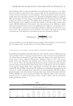
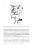
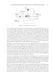
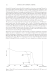
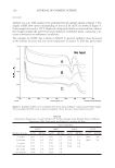
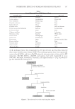
JOURNAL OF COSMETIC SCIENCE 112 practiced to limit contact with the scalp, complaints of discomfort still arise (1,2). It is common for cosmetic products to induce discomfort including itching, stinging, and burning sensations if used incorrectly (1). Although some of these complaints are associ- ated with overuse or misuse, there remains a difference in consumer-perceived discomfort potential between the two most widely used relaxers in the United States. A population-based in vivo study showed that majority of relaxer users perceive no-lye relaxer as less irritating than lye relaxer regardless of whether the discomfort is rated as severe, moderate, or mild (2). Results reported were based on comfort/discomfort evalua- tions of over a thousand salon patrons as an indication of irritation potentials of the two types of relaxers. While reported, these differences are not linked to formulations or bio- chemical processes through scientifi c experimentation. However, there are good reasons to believe that cytokines may be involved as several studies provide evidence of the role of cytokines in inducing sensory discomfort and infl ammation (3–5). Acute infl ammatory pain is characterized by hypernociception due to sensitization of primary sensory neurons. Furthermore, it has been shown that specifi c primary cytokines are released after tissue injury to act on membrane sensory receptors to trigger sensory activation. The nature and type of mediators released depend on the nature of tissue injury. Chen et al. (6) have dem- onstrated the mechanical sensitization of cutaneous C-fi ber nociceptors by prostaglandin E2 (PGE2) in the rat, which provides the basis for studying its possible role in relaxer- induced discomfort. However, no comparative profi ling of cytokines responsible for these perceived differences between lye and no-lye relaxers has been done to date. In this investigation, we used a well-developed three-dimensional reconstructed human epidermis (EpiSkin™ Figure 1) to examine the types and quantity of cytokines associated with sensory irritation that may provide a partial explanation for the perceived differences in discomfort between lye and no-lye relaxers. The cytokines selected in this study are those suggested and confi rmed by many authors (7–9) as being good predictive indicators in both short- and long-term exposure response by the epidermis. MATERIALS AND METHODS MATERIALS Two se parate lots of EpiSkin™ model (1.07 cm2) supplied as a 12-well kit and all neces- sary accessories were purchased from SkinEthic Laboratories (Lyon, France). Upon arrival, tissue inserts were removed from agar and placed in 12-well culture plate containing 2 ml of maintenance medium. Transferred tissues were incubated at 37oC, 5% CO2, and 95% relative humidity for a period of 24 h before using for all experiments. Consumer lye (Mizani, Chicago, IL) and no-lye (Optimum Care, New York, NY) relaxers were pur- chased and used according to instructions in the product insert. PRODUCT APPLICATION Separate EpiSkin™ tissues were topically exposed to75 μl each of medium strength lye relaxer or no-lye relaxer for exactly 15 min. Treated tissues were washed three times, each

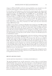
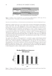
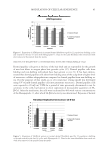
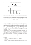
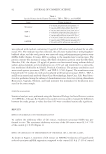
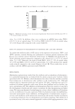
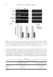
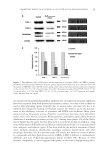
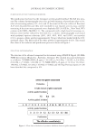
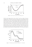
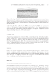
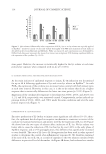
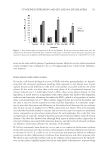
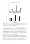
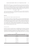
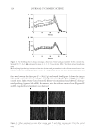
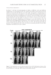
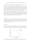
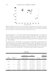
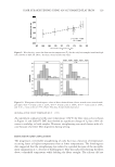
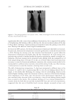
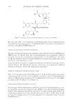
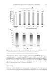
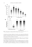
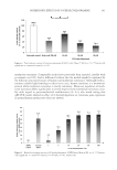
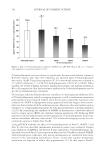
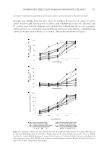
CYTOKINES EXPRESSION AND LYE AND NO-LYE RELAXERS 113 with 25 ml phosphate-buffered saline (PBS) containing Ca++ and Mg++ and incubated at 37oC, 5% CO2, and 95% relative humidity for a period of 4, 24, or 48 h. Cytokines were extracted from the tissues into the media by shaking at 300 rpm for 15 min, and collected aliquots were kept frozen at −20°C until assayed. CYTOKINE ASSAY A panel of proinfl ammatory cytokines [PGE2, interleukin-1α (IL-1α)] and antiinfl am- matory cytokine [IL-1 receptor antagonist (IL-1ra)] in a 2-plex kit was purchased from Millipore Corporation (Billerica, MA). The ELISA kit for PGE2 was obtained from the R&D Systems (Minneapolis, MN). The concentration of mediators was determined by using a Luminex-based multiplex assay system (Luminex Corp, Austin, TX). All assays were performed by following manufacturer’s instructions. STATISTICS Each of two separate experiments using a skin model with different lot number was per- formed in triplicate, and all measurements were in duplicate. The student’s t-test was used to determine signifi cant difference where p ≤0.05 was considered signifi cant. Results were calculated as a difference between the normalized control (100%) and observed value at each time period and are given as mean ± SE. RESULTS LYE AND NO-LYE RELAXERS DIFFERENTIALLY EXPRESS PGE2 Prostaglandins are produced as by-products of arachidonic acid metabolism and are known to induce sensory discomfort in humans and animals (4,6). To explore the possible involvement of the cytokine in the differential sensory perception of discomfort between lye and no-lye relaxers, its expression was examined. As shown in Figure 2, both relaxers induce a statistically signifi cant higher level of PGE2 compared to the control at each Figure 1. Histology of EpiSkin™ showing epidermal structures. A cross section of reconstructed human epidermis stained with hematoxylin and eosin. The model consists of human-derived epidermal keratinocytes that have been grown on bovine collagen coated with collagens 1 and 5. Cultures are raised in air interface to form a multilayered structure of stratum corneum (SC), stratum granulosum (SG), stratum spinosum (SS), and basal layer (SB).
Purchased for the exclusive use of nofirst nolast (unknown)
From: SCC Media Library & Resource Center (library.scconline.org)