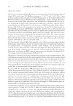
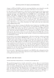
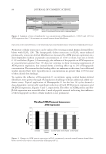
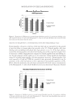
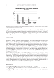
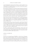
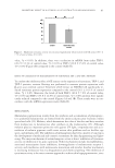
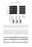
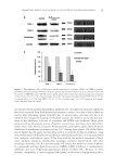
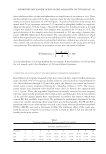
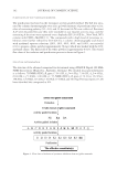
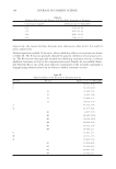
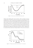
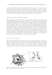
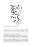
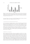
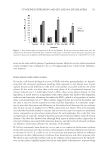
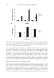
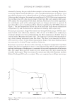
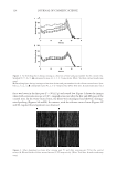
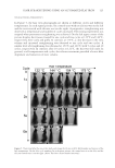
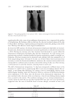
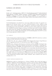
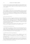
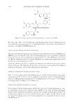
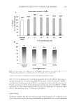
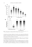
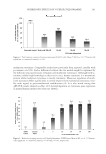
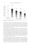
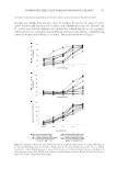
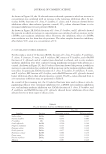
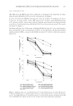
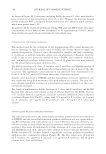
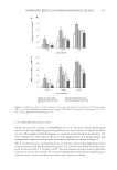
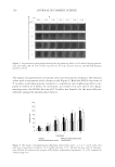
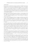
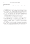
JOURNAL OF COSMETIC SCIENCE 114 time point. However, the increase is statistically higher for the lye relaxer at each time period after exposure when compared with no-lye (p 0.001). IL-1α INDUCTION LEVELS ARE INFLUENCED BY RELAXER TYPE As the main initiator of epidermal response to injury, IL-1α induction was determined for up to 48 h following application of lye and or no-lye relaxer on EpiSkin™. As with PGE2, the expression of the cytokine was increased versus control with both relaxer types at each time interval. However, in this case, it is the no-lye relaxer that elicits a higher response that is statistically different at the latter two time points (p 0.01) (Figure 3). Expression of the cytokine after exposure to lye relaxer was 368%, 256%, and 201% over 4, 24, and 48 h, respectively, over untreated control. Comparatively, no-lye induced ex- pression levels of 436%, 468%, and 338% under the same conditions and over the same period, respectively (Figure 3). EXPRESSION OF IL-1ra BY RELAXERS Excessive production of IL-1α due to injury causes signifi cant side effects (10–13) there- fore, the epidermis has developed an exquisite mechanism to counteract activities of the cytokine to maintain homeostasis and protect itself by the expression of IL-1ra (8). Figure 4A shows how the two types of relaxers affect induction of IL-1ra. No-lye relaxer was better able to cause the induction of IL-1ra than lye by over 140% in the early phase of EpiSkin response, and at 24 h postapplication, this difference has signifi cantly increased to over fourfold. The ratio of IL-1ra to IL-1α expression has been used in other reported studies to determine the control of an infl ammatory response (8). We therefore examined if the ratio of IL-1ra to IL-1α, as a result of exposure to the two relaxers correlated with differences in perceived level of discomfort. As Figure 4B shows, both relaxers have similar Figure 2. Hair relaxers differentially induce expression of PGE2. Lye or no-lye relaxer was topically applied on EpiSkin™ model for exactly 15 min and washed thoroughly with PBS and incubated in fresh media as described in the section Materials and Methods. PGE2 was extracted, and concentration was determined by ELISA. Each data point represents the mean of six tissues run in duplicate ± SE showing signifi cant differen- tial expression of the cytokine (*p 0.05).

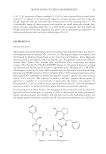
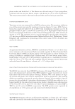
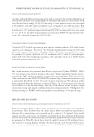
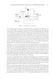
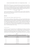
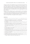
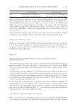
CYTOKINES EXPRESSION AND LYE AND NO-LYE RELAXERS 115 ratios in the early and late phases of epidermal response. However, no-lye relaxer treatment results in higher ratio compared to lye at 24 h suggesting better control of the infl amma- tory response. DISCUSSION AND CONCLUSION Given the well-known biological activity of PGE2 and other prostaglandins, we hypoth- esize that the increased expression resulting from exposure to lye relaxer may in part explain the perceived difference in the level of discomfort associated with the use of the relaxer. In this work, we show that in the early phase (4 h) of epidermal response, lye relaxer induced over 1.5-fold higher level in PGE2 than no-lye relaxer (Figure 2). Our hypothesis as stated above is in agreement with other authors that showed dose-dependent induction and intensity of sensory discomfort by PGE2, which is a by-product of arachi- donic acid metabolism (14–17). These authors suggested that the level of discomfort and the time it takes to feel the sensation are time and dose dependent. It is therefore tempt- ing to speculate that perceived differences in discomfort levels between the two relaxers may be due in part to higher level of PGE2 induced by lye relaxer. This level probably exceeded the threshold needed to saturate and activate nociceptive receptors on peripheral nerves compared to no-lye relaxer induced level. To our knowledge, this is the fi rst time an expression of a specifi c mediator has been associated with discomfort induced by hair relaxers. Our data also showed that although both types of relaxers induce expression of selected cytokines associated with irritation, kinetics and the levels of released cytokines are different, which may infl uence the intensity of discomfort. IL-1α has consistently been shown by in vitro and in vivo studies to be the main cytokine produced by keratinocytes that initiates and propagates epidermal infl ammatory response to irritants (5–8,18). It is able to act in an autocrine and paracrine fashion to infl uence production of other infl ammatory cytokines from keratinocytes and other immune cells. We show in our study that both lye and no-lye relaxers induce expression of IL-1α how- ever, no-lye induces slightly higher level in the early phase of keratinocyte response along Figure 3. Post-relaxer induced expression of IL-1α by EpiSkin. IL-1α was extracted from tissue into the medium and determined by ELISA after treatment with relaxer as described. Each data point represents the mean of six tissues run in duplicate ± SE showing increased IL-1α expression by no-lye at 24 and 48 h (*p 0.05).
Purchased for the exclusive use of nofirst nolast (unknown)
From: SCC Media Library & Resource Center (library.scconline.org)