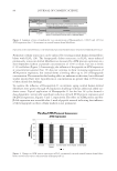
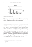
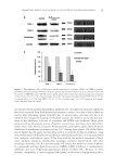
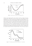
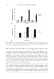
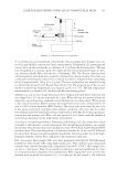
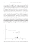
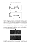
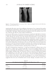
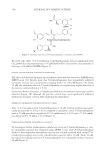
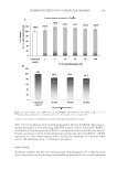
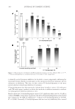
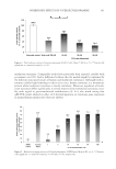
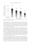
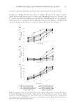
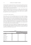
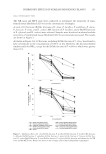
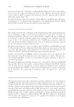
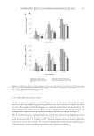
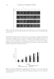
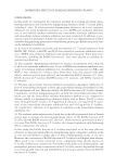
JOURNAL OF COSMETIC SCIENCE 116 with a signifi cant increase of the cytokine at 24 h (Figure 3). As may be expected, no-lye relaxer with a greater ability to induce the expression of IL-1α results in higher produc- tion of IL-1ra. Both relaxer types in fact show increased levels of IL-1ra however, no-lye relaxer elicits a statistically signifi cant higher level at all time intervals. With this result, we show IL-1ra/IL-1α ratio is higher with no-lye than lye relaxer at 24 h (Figure 4B), suggesting it is better able to control the infl ammatory process compared to the lye. However, this difference is absent at 4 and 48 h indicating that both relaxers elicit simi- lar control at those time points. This seems contrary to the fact that the no-lye is reported to be less irritating to the scalp in the early phase. Therefore, one may conclude that the irritation experienced by the users of lye relaxer, which occurs within 20 min of contact is not directly related to the level of IL-1α induced. Both lye and no-lye relaxers are high pH complex formulations so it is not known at this time if a specifi c ingredient or pH is responsible for the perceived phenomenon. A differ- ence in pH may be suspected to infl uence the level of sensory response as it is known that alkaline reagents at pH 12 or higher are corrosive to mammalian tissues. Regular strength lye relaxers have an alkalinity of 0.51 meq/g compared with 0.65 meq/g for regular strength no-lye relaxers. Since no-lye relaxers have higher alkalinity but are perceived to be less irritating, the differences in sensory perception are not likely related to differences Figure 4. Hair relaxers induce expression of IL-1ra. Exposure of EpiSkin™ to no-lye relaxer induced signifi - cantly higher level of IL-1ra compared to lye (A) at all time points. Each data point represents the mean of six tissues run in duplicate ± SE (*p 0.05). (B) IL-ra/IL-1 ratio was determined, which shows that no-lye is better able to control IL-1α–induced acute epidermal response.

CYTOKINES EXPRESSION AND LYE AND NO-LYE RELAXERS 117 in alkalinity. This observation suggests that other mechanisms may be involved in differ- ences in sensory perception between the two relaxers as reported by consumers. It is likely that further studies will be required to explore the role of individual ingredients to an- swer the question. REFERENCES (1) A. N. Syed, Ethnic hair care products, J. Soc. Cosmet. Chem., 12, 235–259 (1995). (2) A. N. Syed and A. R. Naqvi, Comparing the irritation potential of lye and no-lye relaxers, Cosmet. Toiletries, 115, 47–52 (2000). (3) T. M. Cunha, W. A. Verri Jr, J. S. Silva, S. Poole, F. Q. Cunha, and S. H. Ferreira, A cascade of cytokines mediates mechanical hypernociception in mice, Proc. Natl Acad. Sci. USA, 105, 1755–1760 (2005). (4) W. A. Verri Jr, T. M. Cunha, C. A. Parada, S. Poole, F. Q. Cunha, and S. H. Ferreira, Hypernociceptive role of cytokines and chemokines: Targets for analgesic drug development? Pharmacol. Ther., 112, 116–138 (2006). (5) D. Julius and A. I. Bausbaum, Molecular mechanisms of nociception, Nature, 413, 203–210 (2001). (6) X. Chen, K. Tanner, and J. D. Levin, Mechanical sensitization of cutaneous C-fi ber nociceptors by PGE2 in the rat, Neurosci. Lett., 267, 105–108 (1999). (7) G. F. Gerberick and M. K. Robinson, A skin sensitization risk approach for evaluation of new ingredi- ents and products, Am. J. Contact Dermatitis, 11, 65–73 (2000). (8) M. A. Perkins, C. W. Cardin, M. A. Osterheus, M. A. Farage, and M. K. Robinson, A non-invasive method to assess skin irritation and compromised skin conditions using simple tape adsorption of molecular markers of infl ammation, Skin Res. Technol., 7, 227–337 (2001). (9) J. Hauser, H. Saurat, A. Schmitt, F. Jaunin, and J. M. Dayer, Interleukin-1α is present in normal human epidermis, J. Immunol., 136, 3317–3323 (1986). (10) T. S. Kupper and R. W. Groves, The interleukin-1 axis and cutaneous infl ammation, J. Invest. Dermatol., 105, 62s–66s (1995). (11) T. Agner, J. Serup, V. Handlos, and W. Batsberg, Different skin irritation abilities of different qualities of sodium lauryl sulphate, Contact Dermatitis, 21, 184–188 (1989). (12) B. G. Green and J. Bluth, Measuring chemosensory irritability of human skin, J. Cutan. Ocul. Toxicol., 14, 23–48 (1995). (13) D. Veronesi, M. Sailstad, D. L. Doerfl er, and M. Selgrade, Neuropeptide modulation of chemically induced skin irritation, Toxicol. Appl. Pharmacol., 135, 258–267 (1995). (14) T. Minami, H. Nakano, T. Kobayashi, Y. Sugimoto, F. Ushikubi, A. Ichikawa, S. Narumiya, and S. Ito, Characterization of EP receptor subtypes responsible for prostaglandin E2-induced pain responses by use of EP1 and EP3 receptor knockout mice, Br. J. Pharmacol., 133, 438–444 (2001). (15) T. E. Taylor-Clark, B. J. Undem, D. W. MacGlashan Jr, S. Ghatta, M. J. Carr, and M. A. McAlexander, Prostaglandin-induced activation of nociceptive neurons via direct interaction with transient receptor potential A1 (TRPA1), Mol. Pharmacol., 73, 274–281 (2008). (16) O. Hagermark, K. Strandberg, and M. Hamberg, Potentiation of itch and fl are responses in human skin by prostaglandins E2 and H2 and a prostaglandin endoperoxide analog, J. Invest. Dermatol., 69, 527–530 (1977). (17) T. M. Cunha, W. A. Verri Jr, G. G. Vivancos, I. F. Moreira, S. Reis, C. A. Paranda, F. Q. Cunha, and S. H. Ferreira, An electronic pressure-meter nociception paw test for mice, Braz. J. Med. Biol. Res., 37, 401–407 (2004). (18) J. Nickoloff and J. Naidu, Perturbation of epidermal barrier function correlates with irritation of cytokine cascade in human skin, J. Am. Acad. Dermatol., 30, 535–546 (1994).
Purchased for the exclusive use of nofirst nolast (unknown)
From: SCC Media Library & Resource Center (library.scconline.org)