JOURNAL OF COSMETIC SCIENCE 134 In mammalian melanocytes, melanin polymers are produced within specifi c lysosome- related organelles called melanosomes. Melanosomes contain three major pigmentary en- zymes: tyrosinase, tyrosinase-related protein-1, and tyrosinase-related protein-2 (dopachrome tautomerase). By catalyzing the two rate-limiting steps in melanogenesis, i.e., the conver- sion of L -tyrosine to L -3,4-dihydroxyphenylalanine (L-DOPA) and its subsequent oxidation to dopaquinone (2,3), tyrosinase plays a critical role in the melanogenesis process (4). Melanosomes undergo a four-step maturation process characterized by different mor- phological stages (I to IV). As observed using transmission electron microscopy, mela- nosomes in stage I appear as round, clear, unpigmented organelles with intralumenal vesicles. Late stage I melanosomes exhibit proteinaceous fi brils that are completely formed in not yet pigmented melanosomes in stage II. The production of internal ma- trix fi bers as well as the maturation of melanosomes from stages I to II depend on the presence of the structural protein Pmel17, also known as gp100 or SILV. Shortly after its delivery from the trans-Golgi network to stage I melanosomes, Pmel17 is cleaved into several fragments, of which some will form the fi brillar matrix of the organelle (5,6). Pmel17 expression, stability, traffi cking, and processing are principally affected by another melanosomal protein called MART-1 (7). The two proteins form a complex, which suggests that MART-1 acts as a chaperone-like structural component for Pmel17 (8).Once the fi brous striations are fully formed in ellipsoidal stage II melanosomes, tyrosinase is transported to stage III melanosomes, which triggers melanin synthesis. Melanin polymers deposit on the fi brils, resulting in their progressive thickening and darkening. Stage IV of so-called mature melanosomes is fi nally described as the stage where internal structures are no longer distinguishable (9). Development of safe yet effective melanogenesis inhibitors is one of the challenges for the dermatological research and cosmetics industry. Despite numerous chemical steps involving several enzymes, transport, and structural proteins, tyrosinase is considered the rate-limiting enzyme of melanogenesis. As a consequence, the majority of com- mercially available skin-lightening ingredients used over the past decades act—at least in vitro—as tyrosinase inhibitors. Kojic acid (1 0), arbutin (11), licorice extract (12), n-butylresorcinol (13) are among the best known members. However, there are some exceptions such as ascorbic acid and niacinamide (14) that were shown to exert their depigmenting effect through signifi cant antioxidant activity and by inhibiting the transfer of melanosome to keratinocytes, respectively. The aim of our research was to develop a structurally characterized tyrosinase inhibitor devoid of cell toxicity. For this, we chose to focus our investigations on compounds hav- ing high structural homology with the natural substrates of human tyrosinase, namely L -tyrosine and L -DOPA and that would thus act as substrate-mimicking inhibitor. This way, we selected N-feruloyldopamine (also referred to hereafter as N-feruloyldopamine), a naturally occurring ferulic acid derivative as the best candidate. To demonstrate the effi cacy of this molecule at inhibiting melanogenesis, its ability to inhibit mushroom and human tyrosinases in vitro as well as melanin production in vitro was assessed. Furthermore, knowing that antioxidant capacities can downregu- late melanin production, its radical-scavenging property was also evaluated. Finally, its capacity to act on melanosome maturation was also assessed by measuring gene expression of three proteins involved in melanosome formation, namely Pmel17, MART-1, and Protein P.

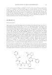
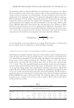
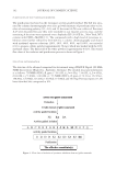
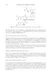
INHIBITORY EFFECTS OF N-FERULOYLDOPAMINE 135 MATERIALS AND METHODS CHEMICALS Ferulic acid, 3-hydroxytyramine, EDCI (N-(3-dimethylaminopropyl)-N′-ethylcarbodiimide hydrochloride), L -DOPA, synthetic melanin, 3-methyl-2-benzothiazolinone hydrazone (MBTH), mushroom tyrosinase, dimethyl sulfoxide (DMSO), and 1,1-diphenyl-2- picrylhydrazyl (DPPH) were purchased from Sigma-Aldrich (Saint-Quentin-Fallavier, France). SYNTHESIS OF N-FERULOYLDOPAMINE N-feruloyldopamine was obtained in a one-step synthesis by means of a peptide cou- pling reaction between ferulic acid and 3-hydroxytyramine in basic medium using EDCI as water soluble coupling agent. Subsequent crystallization affords obtaining N- feruloyldopamine with around 50% yield. The structure of the synthesized compound was confi rmed by nuclear magnetic resonance and mass spectrometry analyses and fur- ther characterized by infrared and high-performance liquid chromatographic (HPLC) analyses (data not shown). The fi nal purity of N-feruloyldopamine was ≥93% as mea- sured using appropriate HPLC method. CELL VIABILITY Cells [normal human epidermal melanocytes (NHEMs) or B16-F10 cells] were seeded at a density of 8000 cells/well in 96-well plates. The B16-F10 cells were cultured in RPMI 1640 medium (Invitrogen, Cergy-Pontoise, France) supplemented with 10% fetal bovine serum (FBS Invitrogen). NHEMs were cultured in keratinocyte serum-free medium (KSFM Invitrogen) sup- plemented with 2% FBS, basic fi broblast growth factor, endothelin-1, α-melanocyte– stimulating hormone (α-MSH), and isoproterenol for 24 h, and then switched to the same medium containing the compounds to test at increasing concentrations and incu- bated for additional 72 h. After incubation, the medium was removed and 0.5 mg/ml MTT (3-(4,5-dimethylthiazol-2-yl)-2,5-diphenyltetrazolium bromide) solution was then added. After 2 h of incubation, the MTT solution was discarded and fi nally DMSO was added. Absorbance [optical density (OD)] was read at 550 nm using Wallac Victor 2 Spectrophotometer (Perkin Elmer, Turku, Finland). Each condition was tested in sextuplicate (n = 6). HUMAN TYROSINASE INHIBITION ASSAY NHEM were seeded in 24-well plates at a density of 80000 cells per well and grown to confl uence. Inhibitors (N-feruloyldopamine or positive references) were diluted in DMSO and next applied in the culture medium for 24 h at 37°C under 5% CO2. After incuba- tion, the culture medium was removed and human tyrosinase was extracted by lysis of melanocytes by thermal shock. After centrifugation, the supernatants containing tyrosinase were incubated with MBTH and L -DOPA solutions for 30 min before the OD was read at 490 nm. Results are expressed as percentage of tyrosinase activity compared
Purchased for the exclusive use of nofirst nolast (unknown)
From: SCC Media Library & Resource Center (library.scconline.org)