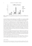
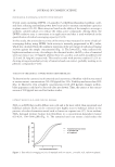
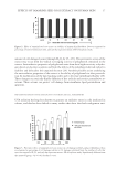
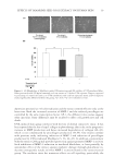
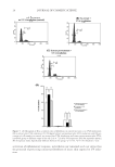
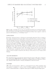
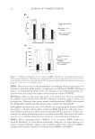
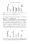
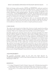
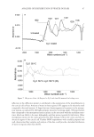
JOURNAL OF COSMETIC SCIENCE 22 DPPH: This assay is based on the measurement of scavenging ability of antioxidant test substances toward the stable radical, 2,2-diphenyl-1-picrylhydrazyl (DPPH). Hydrogen atom or electron-donating ability of the test substances is spectrophotometrically mea- sured from the bleaching of the purple-colored methanol solution of DPPH. Fibroblasts: These are the major cells found in the dermal skin. They are responsible for synthesis of extracellular matrix (ECM) components including collagen, elastin, and proteoglycans. Moreover, they secrete matrix metalloproteinases (MMPs), the enzymes that break down collagen and other proteins that comprise the dermal ECM. G0/G1 phase: In normal skin, some 30% of basal cells are preparing for division. Follow- ing mitosis, a cell enters the G1 phase, synthesizes RNA and protein, and grows in size. Later, DNA is synthesized (S phase) and chromosomal DNA is replicated. A short post- synthetic (G2) phase of further growth occurs before mitosis (M). Some basal cells remain inactive in a so-called G0 phase but may reenter the cycle and resume proliferation. MMP-1: Matrix metalloproteinase-1 (MMP-1) is one of catalytic MMPs enzyme pro- duced by fi broblasts. It is also known as fi broblast collagenase. Its major function is to initiate cleavage of fi brillar collagen (type I and III in skin). Once cleaved by MMP-1, collagen can be further degraded by elevated levels of MMP-3 and MMP-9. Figure 7. (A) Effects of tamarind seed coat extract on MMP-1 and (B) type I procollagen production by UVA-irradiated fi broblasts. Fibroblasts were pretreated with 200 μg/ml tamarind seed coat extract for 24 h before irradiation. Each bar represents mean ± SD of triplicate study. Signifi cantly different between two group *p 0.05, **p 0.01 (Student’s t-test).

EFFECTS OF TAMARIND SEED COAT EXTRACT ON HUMAN SKIN 23 REFERENCES (1) D. He, J. Sun, X. Zhu, S. Nian, and J. Liu, Compound K increases type I procollagen level and decreases matrix metalloproteinase-1 activity and level in ultraviolet-A-irradiated fi broblasts, J. Formos. Med. Assoc., 110, 153–160 (2011). (2) N. Buechner, P. Schroeder, S. Jakob, K. Kunze, T. Maresch, C. Calles, J. Krutmann, and J. Haendeler, Changes of MMP-1 and collagen type Iα1 by UVA, UVB and IRA are differentially regulated by Trx-1, Exp. Gerontol., 43, 633–637 (2008). (3) H. Watanabe, T. Shimizu, J. Nishihira, R. Abe, T. Nakayama, M. Taniguchi, H. Sabe, T. Ishibashi, and H. Shimizu, Ultraviolet A-induced production of matrix metalloproteinase-1 is mediated by macrophage migration inhibitory factor (MIF) in human dermal fi broblasts, J. Biol. Chem., 279, 1676–1683 (2004). (4) G. Jenkins, Molecular mechanisms of skin ageing, Mech. Ageing Dev., 123, 801–810 (2002). (5) E. C. Naylor, R. E. B. Watson, and M. J. Sherratt, Molecular aspects of skin aging, Maturitas, 69, 249–256 (2011). (6) T. M. Hagen, Oxidative stress, redox imbalance, and the aging process, Antioxid. Redox. Signal., 5, 503–506 (2003). (7) P.-J. Chua, G. W.-C. Yip, and B.-H. Bay, Cell cycle arrest induced by hydrogen peroxide is associated with modulation of oxidative stress related genes in breast cancer cells, Exp. Biol. Med., 234, 1086–1094 (2009). (8) M. Li, L. Zhao, J. Liu, A.-L. Liu, W.-S. Zeng, S.-Q. Luo, and X.-C. Bai, Hydrogen peroxide induces G2 cell cycle arrest and inhibits cell proliferation in osteoblasts, Anat. Rec., 292, 1107–1113 (2009). (9) F. Hazane, S. Sauvaigo, T. Douki, A. Favier, and J.-C. Beani, Age-dependent DNA repair and cell cycle distribution of human skin fibroblasts in response to UVA irradiation, J. Phytochem. Photobiol., 82, 214– 223 (2006). (10) T. Pluemsamran, T. Onkoksoong, and U. Panich, Caffeic acid and ferulic acid inhibit UVA-induced matrix metalloproteinase-1 through regulation of antioxidant defense system in keratinocyte HaCaT cells, Photochem. Photobiol., 88, 961–968 (2012). (11) J. Viyoch, S. Buranajaree, F. Grandmottet, S. Robin, D. Binda, C. Viennet, N. Waranuch, and P. Humbert, Evaluation of the effect of Thai breadfruit’s heartwood extract on the biological functions of fi broblasts from wrinkles, J. Cosmet. Sci., 61, 311–324 (2010). (12) J. Kim, J. S. Hwang, Y. K. Cho, Y. Han, Y. J. Jeon, and K. H. Yang, Protective effects of (-)-epigallocatechin- 3-gallate on UVA- and UVB-induced skin damage, Skin Pharmacol. Appl. Skin Physiol., 14, 11–19 (2001). (13) G. Pumthong, Antioxidative activity of polyphenolic compounds extracted from seed coat of Tamarindus indicus Linn. PhD Thesis, Chiangmai Mai University, Thailand, 1999. (14) T. Tsuda, M. Watanabe, K. Ohshima, A. Yamamoto, S. Kawakishi, and T. Osawa, Antioxidative components isolated from the seed of tamarind (Tamarindus indica L.), J. Agric. Food Chem., 42, 2671–2674 (1994). (15) W. Aengwanich, M. Suttajit, T. Srikhun, and T. Boonsorn, Antibiotic effect of polyphenolic compound extracted from tamarind (Tamarindus indica L.) seed coat on productive performance of broilers, Int. J. Poult. Sci., 8, 749–751 (2009). (16) T. Komutarin, S. Azadi, L. Butterworth, D. Keil, B. Chitsomboon, M. Suttajit, and B. J. Meade, Extract of the seed coat of Tamarindus indica inhibits nitric oxide product by murine macrophages in vitro and in vivo, Food Chem. Toxicol., 42, 649–658 (2004). (17) S. Intaraprasit, A. Faikrua, A. Sittichokechaiwut, and J. Viyoch, Effi cacy evaluation of the fi broblast- seeded collagen/chitosan scaffold on application in skin tissue engineering, ScienceAsia, 38, 268–277 (2012). (18) P. Inpanya, A. Faikrua, A. Ounaroon, A. Sittichokechaiwut, and J. Viyoch, Effects of the blended fi - broin/aloe gel fi lm on wound healing in streptozotocin-induced diabetic rats, Biomed. Mater., 7, 035008 (2012). doi: 10.1088/1748-6041/7/3/035008. (19) K. Tanaka, N. Fujita, M. Yoshioka, and N. Ogawa, Immunosuppressive and non-immunosuppressive immunophilin ligands improve H2O2-induced cell damage by increasing glutathione levels in NG108- 15 cells, Brain Res., 889, 225–228 (2001). (20) M. I. Gil, F. A. Tomás-Barberán, B. Hess-Pierce, and A. A. Kader, Antioxidant capacities, phenolic compounds, carotenoids, and vitamin C contents of nectarine, peach, and plum cultivars from Califor- nia, J. Agric. Food Chem., 50, 4976–4982 (2002). (21) L. P. Leong and G. Shui, An investigation of antioxidant capacity of fruits in Singapore markets, Food Chem., 76, 69–75 (2002). (22) C. Guo, J. Yang, J. Wei, Y. Li, J. Xu, and Y. Jiang, Antioxidant activities of peel, pulp and seed fractions of common fruits as determined by FRAP assay, Nutr. Res., 23, 1719–1726 (2003).
Purchased for the exclusive use of nofirst nolast (unknown)
From: SCC Media Library & Resource Center (library.scconline.org)