JOURNAL OF COSMETIC SCIENCE 24 (23) A. Floegel, D.-O. Kim, S.-J. Chung, S. I. Koo, and O. K. Chun, Comparison of ABTS/DPPH assays to measure antioxidant capacity in popular antioxidant-rich US foods, J. Food Compos. Anal., 24, 1043– 1048 (2011). (24) J. Deng, W. Cheng, and G. Yang, A novel antioxidant index (AAU) for natural products using the DPPH assay, Food Chem., 125, 1430–1435 (2011). (25) N. Lourith, M. Kanlayavattanakul, and S. Chanpirom, Free radical scavenging activity of tamarind seed coat and its cosmetic application, J. Health Res., 23, 159–162 (2009). (26) Y. Sudjaroen, R. Haubner, G. Würtele, W. E. Hull, G. Erben, B. Spiegelhalder, S. Changbumrung, H. Bartsch, and R. W. Owen, Isolation and structure elucidation of phenolic antioxidants from tamarind (Tamarindus indica L.) seeds and pericarp, Food Chem. Toxicol., 43, 1673–82 (2005). (27) W. Dröge, Free radicals in the physiological control of cell function, Physiol. Rev., 82, 47–95 (2002). (28) C. A. Rice-Evans and N. J. Miller, Antioxidant activities of fl avonoids as bioactive components of food, Biochem. Soc. Trans., 24, 790–795(1996). (29) A. Arora, T. M. Byrem, M. G. Nair, and G. M. Strasburg, Modulation of liposomal membrane fl uidity by fl avonoids and isofl avonoids, Arch. Biochem. Biophys., 373, 102–109 (2000). (30) A. Tarozzi, A. Marchesi, S. Hrelia, C. Angeloni, V. Andrisano, J. Fiori, G. Cantelli-Forti, and P. Hrelia, Protective effects of cyanidin-3-O-β-glucopyranoside against UVA-induced oxidative stress in human keratinocytes, Photochem. Photobiol., 81, 623–629 (2005). (31) R. P. Singh, S. Sharad, and S. Kapur, Free radicals and oxidative stress in neurodegenerative diseases: Relevance of dietary antioxidants, J. Indian Acad. Clin. Med., 5, 218–225 (2004). (32) A. Chiu and A. B. Kimball, Topical vitamins, minerals and botanical ingredients as modulators of en- vironmental and chronological skin damage, Br. J. Dermatol., 149, 681–691 (2003). (33) A. Svobodová, J. Psotová, and D. Walterová, Natural phenolics in the prevention of UV-induced skin damage. A review, Biomed. Pap., 147, 137–145 (2003). (34) Y. Y. He, J. L. Huang, D. C. Ramirez, and C. F. Chignell, Role of reduced glutathione effl ux in apopto- sis of immortalized human keratinocytes induced by UVA, J. Biol. Chem, 278, 8058–8064 (2003). (35) L. Yin, A. Morita, and T. Tsuji, Skin aging induced by ultraviolet exposure and tobacco smoking: Evi- dence from epidemiological and molecular studies, Photodermatol. Photoimmunol. Photomed., 17, 178–183 (2001). (36) M. Wlaschek, G. Heinen, A. Poswig, A. Schwarz, T. Krieg, and K. Scharffetter-Kochanek, UVA-induced autocrine stimulation of fi broblast-derived collagenase/MMP-1 by interrelated loops of interleukin-1 and interleukin-6, Photochem. Photobiol., 59, 550–556 (1994). (37) Z. Liu, H. Chen, H. Yang, J. Liang, and X. Li, Low-dose UVA radiation-induced adaptive response in cultured human dermal fi broblasts, Int. J. Photoenergy 2012, 1–11 (2012). (38) F. Afaq and H. Mukhtar, Effects of solar radiation on cutaneous detoxification pathways, J. Photochem. Photobiol. B., 63, 61–69 (2001). (39) C. L. Hammond, T. K. Lee, and N. Ballatori, Novel roles for glutathione in gene expression, cell death, and membrane transport of organic solutes, J. Hepatol., 34, 946–954 (2001). (40) T. Quan, Z. Qin, W. Xia, Y. Shao, J. J. Voorhees, and G. J. Fisher, Matrix-degrading metalloproteinases in photoaging, J. Investig. Dermatol. Symp. Proc., 14, 20–24 (2009). (41) M. Brennan, H. Bhatti, K. C. Nerusu, N. Bhagavathula, S. Kang, G. J. Fisher, J. Varani, and J. J. Voorhees, Matrix mettalloproteinase-1 is the major collagenolytic enzyme responsible for collagen damage in UV- irradiated human skin, Photochem. Photobiol., 78, 43–48 (2003). (42) G. J. Fisher, H. C. Choi, Z. Bata-Csorgo, Y. Shao, S. Datta, Z. Q. Wang, S. Kang, and J. J. Voorhees, Ultraviolet irradiation increases matrix metalloproteinase-8 protein in human skin in vivo, J. Invest. Dermatol., 117, 219–226 (2001). (43) T. Quan, T. He, S. Kang, J. J. Voorhees, and G. J. Fisher, Ultraviolet irradiation alters transforming growth factor beta/smad pathway in human skin in vivo, J. Invest. Dermatol., 119, 499–506 (2002). (44) L. Yin, A. Morita, and T. Tsuji, The crucial role of TGF-b in the age-related alterations induced by ul- traviolet A irradiation, J. Invest. Dermatol., 120, 703–705 (2003). (45) X. Z. Song, J. P. Xia, and Z. G. Bi, Effects of (-)-epigallocatechin-3-gallate on expression of matrix metalloproteinase-1 and tissue inhibitor of metalloproteinase-1 in fi broblasts irradiated with ultraviolet A, Chin. Med. J. (Engl.), 117, 1838–1841 (2004).

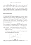
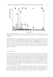
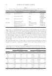
J. Cosmet. Sci., 65, 25–36 (January/February 2014) 25 Molecular marker approaches for tracking redox damage and protection in keratins JOLON M. DYER, CHARISA D. CORNELLISON, ANITA J. GROSVENOR, STEFAN CLERENS, and SANTANU DEB-CHOUDHURY, Food & Bio-Based Products, AgResearch, Christchurch 8140, New Zealand (J.D., C.C., A.G., S.C., S.D.), Riddet Institute at Massey University, Palmerston North, New Zealand (J.D.), and Biomolecular Interaction Centre, University of Canterbury, Christchurch 8140, New Zealand (J.D.). Accepted for publication September 1, 2013. Synopsis There is increasing awareness of the importance of reductive and oxidative (redox) protein damage in protein- based materials including, hair, wool, nails, and skin. Light-induced damage to protein-based materials is of particular concern because of its impact on age-related degradation and product life spans. Consequently, cosmetic applications frequently target hair and skin restoration, where the integrity of the constituent fi la- mentous proteins is essential to a healthy appearance. The keratins constitute an important subset of the structural proteins within skin, hair, and wool. We will introduce a means to assess damage to this important group of proteins at the molecular level, utilizing proteomic techniques to track the formation or degradation of sensitive peptides within intermediate fi lament proteins. The degradation of three molecular markers of redox damage, the peptides SFGYR, LASDDFR, and DVEEWYIR, along with the formation of their oxi- dized products, is demonstrated after exposure to ultraviolet A, ultraviolet B, and blue light. The method is shown to be suitable for evaluating the protective effect of treatments, as lower levels of oxidative markers were observed after the application of a protective fi ber treatment. Molecular-level redox tracking will allow more targeted design and evaluation of protection and repair treatments for protein systems. INTRODUCTION Keratins, previously also referred to as intermediate fi lament proteins, represent an im- portant class of structural proteins of critical relevance to the physico-mechanical proper- ties of mammalian fi bers. Keratins are directly responsible for the structural integrity and associated physical and mechanical properties of hair, wool, skin, and nails (1,2). Both oxidative and reductive damages to these proteins result in deleterious effects, such as loss of strength and an increase in brittleness (3). Address all correspondence to Jolon M. Dyer at jolon.dyer@agresearch.co.nz .
Purchased for the exclusive use of nofirst nolast (unknown)
From: SCC Media Library & Resource Center (library.scconline.org)