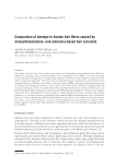
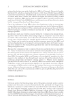
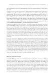
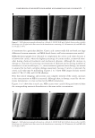
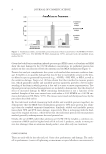
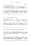
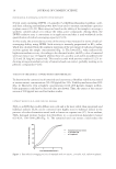
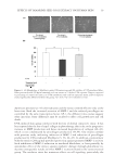
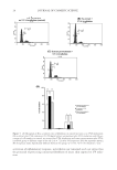
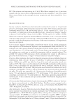
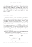
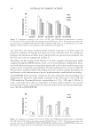
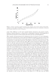
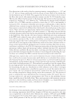
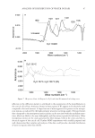
JOURNAL OF COSMETIC SCIENCE 28 a fl ow rate of 8 μl/min. The trap column was then switched in line with the analytical column (C18, 30 cm, 75 μm ID). Both trap column and analytical column were in-house packed with Varian Microsorb C18 5 μm, 300 Å particles. Reversed-phase gradients were from 2% to 55% solvent B at a fl ow rate of 150 nl/min over 50 min. Solvent A was 0.2% formic acid in HPLC-grade water solvent B was 0.2% formic acid in LCMS-grade aceto- nitrile. Loading and desalting solvent was 0.2% formic acid, 2% acetonitrile in HPLC- grade water. The analytical column outlet was directly connected to the mass spectrometer using a stainless steel nanospray needle (Proxeon). Triplicate technical repeats were per- formed for all samples. RESULTS AND DISCUSSION SELECTION OF MOLECULAR MARKERS To characterize redox modifi cation and select marker peptides for oxidative damage, a simplifi ed system of enriched wool proteins was fi rst evaluated. Type I and II keratins were isolated and purifi ed from whole wool utilizing reductive sulfi tolysis (12). Solutions of these keratins were irradiated with UVA for periods between 0 and 72 h. The photo- modifi ed proteins thus produced were enzymatically digested into peptide mixtures. Chromatographic analysis indicated substantial photomodifi cation of the wool-derived proteins with increasing irradiation times, as demonstrated in Figure 1 (peptide oxida- tion is expected to result in an increase in UV absorbance due to the formation of UV- absorbing chromophores (13,14). Electrospray ionization tandem mass spectrometry (ESI-MS/MS) was utilized to search for sequence-modifi ed keratin-derived peptides and to characterize modifi cations correspond- ing to redox-modifi ed residues. Certain residues, particularly the aromatic amino acid resi- dues, were expected to be sensitive to UVA and UVB irradiation through type I and type II photooxidative mechanisms (15–17). From this evaluation, marker peptides for damage tracking were selected. The criteria utilized for the selection of markers were as follows: • The peptide contained a photosensitive aromatic residue: phenylalanine, tyrosine, tryptophan, and/or histidine. • The peptide was reproducibly observed in ESI-MS analysis of the unfractionated keratin tryptic digests with a high relative ion intensity. • The peptide ion m/z and the m/z of corresponding major photoproduct ions were clear of interfering ions in the unfractionated analysis. Figure 1. HPLC traces of keratin digest solutions corresponding to UVA irradiation times of 0, 3, 24, and 72 h (monitored at 214 nm).

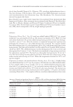
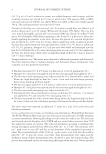
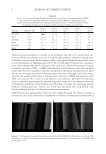
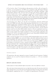
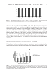
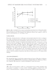
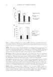
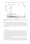
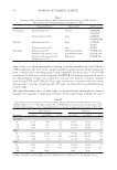
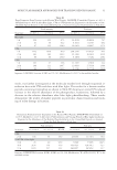
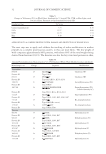
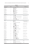
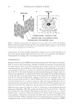
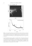
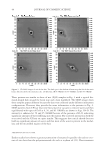
MOLECULAR MARKER APPROACHES FOR TRACKING REDOX DAMAGE 29 An ESI-MS/MS spectrum showing tyrosine photooxidation in the keratin-derived pep- tide with native sequence SFGYR is shown in Figure 2. This peptide met the criteria for use as a molecular marker of redox damage. Using MS/MS fragmentation data, a range of photo-induced redox modifications were thus characterized and located within wool keratins. Those peptides meeting the criteria for potential utilization as redox damage markers are listed in Table I, along with observed UVA-induced photomodifi cations. DAMAGE TRACKING To validate redox damage tracking for keratins, the relative abundance of selected oxida- tive modifi cations to keratin marker peptides was evaluated over differing irradiation protocols. Parallel quantitative evaluation of both the ion intensity and the ion peak area of each oxidative damage product was performed relative to the unmodifi ed native pep- tide within each sample, with each sample analyzed in triplicate. The two differing quan- titative methods were employed in parallel both for comparison purposes and to demonstrate the validity of the marker peptide approach for both methods. This is sum- marized for two peptides identifi ed as good candidates for use as molecular redox damage markers in Tables II and III. In the keratin marker peptide SFGYR, blue light produced an initial increase in the relative abundance of the tyrosine primary oxidation products, dopa, likely due to the UV component of the blue light utilized (11), but this was subsequently photo- bleached by further irradiation, consistent with the absorption of blue light (i.e., Figure 2. ESI-MS/MS spectrum of the keratin-derived peptide SFGY*R (m/z 645.3), where tyrosine (Y) has been photomodifi ed by UVA exposure to dihydroxyphenylalanine (dopa) (Y*), with selected fragment ions highlighted.
Purchased for the exclusive use of nofirst nolast (unknown)
From: SCC Media Library & Resource Center (library.scconline.org)