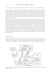
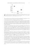
JOURNAL OF COSMETIC SCIENCE 184 neutral lipids which are synthesized de novo by sebocytes. Immature sebocytes are located at the periphery of sebaceous glands and are fl attened cells without lipids. When sebocytes progress to the center of the gland, they mature and increase in size due to lipid synthesis. Ultimately, as the cells differentiate, they disintegrate and release their lipid content into the infundibulum. Human sebum contains cholesterol (1.5–2.5%), cholesterol esters (3–6%), squalene (12–20%), free fatty acids (15–30%), triglycerides (30–50%), and wax esters (26–30%) (2). Overproduction of sebum is a common scheme for teenagers all around the world how- ever, it is also widely distributed in the general population whatever the age, the gender, and the geographical zone. It makes the skin look oily and shiny and renders people less attractive in addition, it creates conditions for the development of infl amed cutaneous alterations (3). Balance of lipids, such as linoleic and arachidonic acids, is usually modi- fi ed these acids act directly or indirectly on lipid storage increase through Peroxisome Proliferator Activated Receptors (PPARs) and perilipin expression. They also trigger pro- duction of infl ammatory mediators such as interleukins (IL-6 and IL-8) and prostaglandin E2 (PGE-2) (4, 5). These mediators, among others, are key players in the increase of lipid synthesis by sebocytes, in the proliferation of microorganism such as Propionibacterium acnes, in keratinocyte hyper-proliferation, and in macrophage invasion of nearby tissues. The lack of an ideal animal model comparable to human sebaceous glands was surmounted by the development of experimental models such as SEB-1, SEB-E6E7, and SZ95 cell lines all immortalized by SV40 (6,7) or using extracted human sebaceous glands. Growth of primary normal human sebocytes (NHSs) in monolayer was achieved by various teams however, these models could not be multiplied because cells loose quickly their features. So, this model remains a challenge in order to evaluate molecules with a nontransformed cell. This paper relates use of NHS to study modulators of lipid storage and variation of pro- infl ammatory mediator synthesis within these cells. Among products that were evalu- ated, a plant cell culture extract of Syringa vulgaris (CCSV) showed signifi cant effects on both lipid storage and on bacterial-induced pro-infl ammatory mediator release. Comple- mentary tests performed on NHK, macrophages, and skin explants confi rmed the interest of CCSV for reduction of PGE-2, IL-6, and IL-8 induced by lipopolysaccharide (LPS) or P. acnes. Moreover, CCSV was evaluated on volunteers with greasy skin, where it reduced sebum synthesis and consequences of over lipid production (skin blemishes, hyperkeratinization). MATERIALS AND METHODS CELL CULTURE, SKIN EXPLANTS, AND REAGENTS NHS. Mycoplasmas-free NHS cells, derived from facial sebaceous gland (woman, 26 years) and obtained in compliance with the procedures and international standards for donation and collection of human tissue used for cell isolation were used. They were amplifi ed and sub- cultured in appropriate and defi ned cell culture medium containing 2–10% of fetal calf serum (FCS, Gibco®,Thermo Fisher Scientifi c Inc., Waltham, MA). Various cell medium were tested for NHS growth and lipogenesis assays, such as Dulbecco’s Modifi ed Essential Medium (DMEM), Dulbecco’s Modifi ed Essential Medium and Ham’s F-12 Medium (DMEM-F12), Keratinocyte Serum-Free Medium (KSFM), Medium 154 (M154) completed with antibiotics

EVALUATION OF MOLECULES OR EXTRACTS MODULATING SEBORRHEA 185 (100 U/ml penicillin, and 100 mg/ml streptomycin), 50 μg/ml EGF, and FCS. Mycoplasma removal treatment was achieved using Plasmocure® kit (Invivogen®, San Diego, CA) and followed regularly (MycoAlert® mycoplasma detection kit, Lonza®, Basel, Switzer- land). For lipid detection, proliferative NHSs were seeded in 96-well fl at and black bot- tom plate (Becton®, Rungis, France). When the cells reached confl uence, contact between cells and compounds was performed for 24–72 h. Cell number and lipid synthesis were estimated, respectively, using fl uorescein diacetate (FDA) and Red Nile fl uorescent probes (both from Sigma®-Aldrich®, St. Louis, MO) (8). Results were obtained using a fl uores- cent reader (FLUOstar®, BMGLabteck®, Ortenberg, Germany) equipped with appropri- ate fi lters. For lipid in situ labelling, cells were fi xed with Histochoice® (Clinisciences®, Nanterre, France) then incubated with Oil Red O® (Sigma®) solution and observed under microscope. For protein in situ labelling, cells were fi xed with Histochoice® then permeabi- lized with 0.1% Triton® X-100 (Sigma-Aldrich®) in phosphate-buffered saline and blocked in 1% bovine serum albumin/0.05% Tween® 20 (Sigma-Aldrich®). Primary antibodies were as follows: anti-K7, anti-K4, anti-fi laggrine, anti-involucrine, and anti-PLIN-2 (Santa Cruz Biotechnology®, Eurogentec®, Dallas, TX) (1). Species-specifi c secondary antibodies were conjugated to Alexa-Fluor® 488 (Life Technologies®, Liège, Belgium). Nuclei were counterstained with propidium iodide or Hoechst dye (Sigma-Aldrich®, Carlsbad, CA). For pro-infl ammatory studies, cells received for 24 h both Escherichia coli LPS (Aldrich®) and CCSV (IRB®, Altavilla Vicentina, Italy) for 3 days. Release of PGE-2 was evaluated using EIA assay (Cayman®). AntiMicrobial Peptide (AMP) cathelicidin and beta-defensin-2 (hBD2) were measured after 3 days from cell lysates using ELISA assays (Hycult Biotech®, Peprotech®, Peprotech, Rocky Hill, NJ). Results were normalized with cells quantifi ca- tion through Hoescht 33258 (Uden, The Netherlands) method. Collagen lattices were pre- pared according to the method of Bell et al. (9). The collagen solution and normal human dermal fi broblast (NHDF), or NHS suspensions, were blended simultaneously for lattice formation. NHDF medium was DMEM with penicillin, streptomycin, and glutamine (DMEMc), whereas NHSs were in their medium, both containing FCS (10%). The lattice diameter was measured every day. Equivalent skins were prepared according to the method of Carlson et al. (10). NHS or NHK were seeded onto NHDF containing lattice in order to evaluate their respective capacities of formation of an epidermis with its stratum cor- neum. All studies were performed at 37°C in 5% CO2. Normal human keratinocytes (NHKs, CELLnTEC®, Bern, Switzerland) in KSFM with Bovine Pituitary Extract and 2.5 μg/ml Epidermal Growth Factor (EGF) (Gibco®) were seeded in plastic vessel until confl uence was reached. Pro-infl ammatory studies were per- formed as mentioned earlier. Release of PGE-2, IL-6, and IL-8 was measured in cell culture supernatants using Enzyme ImmunoAssay (EIA)/ELISA assays (Cayman® Cayman Chem- ical, Ann Arbor, MI) and Pelikine®, Sanquin, Amsterdam, The Netherlands) results were normalized as mentioned earlier. All studies were performed at 37°C in 5% CO2. For macrophages studies, RAW264.7 murine macrophages were cultivated up to confl u- ence in DMEMc containing 10% FCS. They were treated according to Kim et al. (11) with LPS then with CCSV. The amount of nitrite was measured in cell culture superna- tant using the Griess reagent (Cayman®). All studies were performed at 37°C in 5% CO2. Skin explants (0.5 cm², Biopredic®, Biopredic® International, Saint Grégoire, France) were obtained from abdominal region of a Caucasian woman (52 years). Upon receipt, they were transferred into culture media (MIL305, Biopredic®) and cultivated at 37°C in 5% CO2. For studies, explants were in contact with culture media containing P. acnes
Purchased for the exclusive use of nofirst nolast (unknown)
From: SCC Media Library & Resource Center (library.scconline.org)