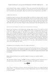
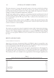
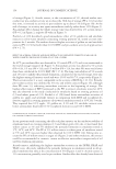
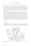
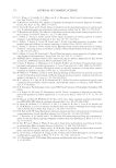
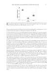
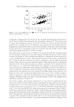
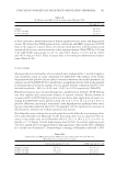
JOURNAL OF COSMETIC SCIENCE 186 inactivated cell extract (Sederma®, Sederma, Le Perray en Yvelines, France), prepared by freezing/thawing cycles. Cream containing 0.8 mM of CCSV (i.e., 0.8 mM phenylpro- panoids) was applied on skin surface a placebo cream was applied as control. After 5 days of a daily topical application, release of PGE-2, IL-6, and IL-8 was measured as men- tioned earlier, or explants were prepared for immunohistochemistry, being frozen and vertically sectioned (7–10 μm) using a cryostat (Leica® CM15105, Leica Biosystems Inc. Buffalo Grove, IL). Presence of Kallikrein-related peptidase 5 (KLK5), formerly known as stratum corneum tryptic enzyme (SCTE), a desquamation enzyme, which en- ables corneodesmosome division and corneocyte release to take place, was evaluated through immunolabelling performed using anti-SCTE antibody (Abcam®, Cambridge, MA). All studies were performed at 37°C in 5% CO2. Statistical analysis values were expressed as means ±SDM of the results for at least three experiments. Anova and Student t test were used for comparisons p values 0.05 were considered statistically signifi cant. IN-VIVO STUDIES Several studies were performed between March 2013 and December 2013 to establish the in vivo effi cacy of CCSV. Each individual study lasted 1 month. For each study and accord- ing to the protocol, volunteers applied by themselves a cream containing 0.8 mM CCSV and/or its vehicle. All the applications were performed on a daily basis in normal condi- tions of use. Each volunteer acted as his own control. Written informed consent was ob- tained from all participants. Medical control and noninvasive methods were used. Clinical studies comply with the latest recommendations of the World Medical Association (Dec- laration of Helsinki, 1964, and its successive updates) and with the French law 2004-806 Figure 1. NHS at passage 7 in NHS-7 medium. Left: 2 days after seeding right: 5 days. ×250. Figure 2. NHS in various culture media with 10% of FCS after 3 days: (A) DMEMc DI3, (B) DMEMc + EGF, (C) KSFM/BPE + EGF, (D) NHS7 Red Oil staining, ×500.

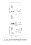
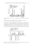
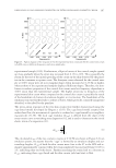
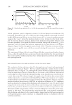
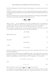
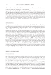
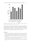
EVALUATION OF MOLECULES OR EXTRACTS MODULATING SEBORRHEA 187 dated August 9, 2004, concerning public health. Moreover, our studies follow the spirit of good clinical practices (ICH E6 GCP, 1996). As a noninterventional study, neither advice of an ethic committee nor submission to competent authorities are required. How- ever, an internal ethic committee was asked to allow these studies. For sebum production study, 23 volunteers (women, [19–32 years] mean: 24.5 years) were recruited, with oily skin and acne prone skin, some regularly had acne-type blem- ishes on the face. Cream containing 0.8 mM CCSV was applied daily onto hemiface, whereas the other hemiface received the corresponding placebo cream. Modulation of sebum production and number of active glands were evaluated using Sebufi x® (Courage & Khazaka®) and image analysis through Mountains Map® software (Digital Surf®). Mea- surements were done one night after the last application. Skin blemishes were evaluated on several panels (Sederma®, Cosmetest®) due to the dif- fi culty in recruiting volunteers with blemishes in a suffi cient number and intensity. Cream applications were the same as the precedent test. Blemishes were evaluated on standardized pictures made under cross polarized lighting with a photographic bench (Orion Concept®). The skin specifi c software Framescan® (Orion Concept®) was used to quantify blemishes based on redness threshold. Hyperkeratosis was performed at BioEC® using cyanoacrylate biopsies on volunteers with oily skin and comedones on face (women and men, [18–45 years] mean: 32 years) after a daily application of a cream containing CCSV. Assessment was achieved after Figure 3. NHS in NHS-7 culture medium + linoleic acid 10-4 M at passage 8 (left picture) and 20 (right picture). Lipids are labeled using Red Oil ×250. Figure 4. NHS in NHS-7 culture medium at passage 9 without (left) or with (middle) linoleic acid 10-4 M. Lipids are labeled using red oil ×250. Right picture: human adipocytes.
Purchased for the exclusive use of nofirst nolast (unknown)
From: SCC Media Library & Resource Center (library.scconline.org)