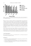
JOURNAL OF COSMETIC SCIENCE 196 diabetic and renal diseases, elevated levels of skin AGEs have been reported in patients with systemic lupus erythematosus (16), chronic cerebral ischemia (17), schizophrenia (18), peripheral artery disease (19,20), and chronic heart failure (21). SAF has also been studied in healthy people, and positive correlations of skin AGE levels with chronological age have been established for Dutch and Slovak Caucasians, Japanese, Chinese, and Saudi Arabians (22–26). Because skin pigmentation can infl uence the results of SAF, corrections for melanin and hemoglobin exist to enable more meaningful correla- tions with age in those individuals with darker skin pigments (27). In the skin, AGEs are considered photosensitizers and can generate reactive oxidative species on ultraviolet (UV) irradiation, which accelerates the skin aging process (28,29). Meanwhile, there have been reports indicating that chronic UVB exposure induces additional fl uorescence exci- tation bands in mice skin (30) and observations of signifi cantly more AGE staining in sun-exposed skin than in sun-protected skin, suggesting that solar irradiation increases dermal glycation (31). Increased skin AGEs has also been correlated with heavy smokers and chronic obstructive pulmonary disease patients (32). Of the SAF studies, most of them has used commercially available noninvasive instru- ments, AGE Reader (DiagnOptics, Groningen, The Netherlands) or SCOUT DS (VeraLight, Albuquerque, NM), to measure the accumulated skin AGEs in vivo. Their detailed oper- ating principles were previously reported (1,8). To date, all studies using SAF as a mea- sure of AGEs have been obtained from the volar forearms except two reports in which the measurements were obtained on the inner aspect of the upper arm skin (23,33). The facial skin has been the primary focus of antiaging research in the skincare industry, and the noninvasive in vivo measurement of facial skin AGEs would add new data to ag- ing research. In this study, we report skin AGEs measured from the left-cheek skin and compare the results with that of the left volar forearm skin. Our aim was to show the site and gender differences of skin AGE level and to correlate the results with participants’ chronological age as well as the level of facial wrinkling. We selected the left cheek for measurement to maximize the effect of sun exposure on the skin because of the driving convention in the United States. MATERIALS AND METHODS SAF MEASUREMENT A commercially available AGE Reader™ SU (DiagnOptics) was used to noninvasively evaluate the level of accumulated AGEs in the skin. Its measurement principle is based on the properties of SAF because the primary components of AGEs in the skin emit a characteristic SAF when excited by UV light. The instrument illuminates a skin surface area of 4 cm2, guarded against surrounding light, with an excitation light source between 300 and 420 nm (peak excitation 370 nm). Emission light and refl ected excitation light from the skin are measured with a spectrometer in the 420–600 nm range. Because skin pigmentation may absorb light and thus infl uence autofl uorescence, skin refl ection mea- surements across the 300–420 nm range were compared with those of a white Tefl on block (1). SAF is calculated as the ratio of the light intensity refl ected by the skin in the 420–600 nm wavelength range and the light intensity in the 300–420 nm wavelength range and is represented as the skin autofl uorescence ratio (AFR) in AGE Reader (1).

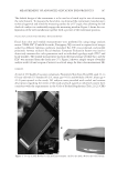
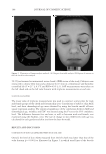
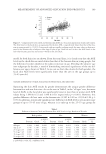
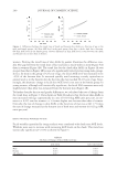
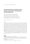
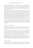
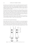
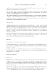
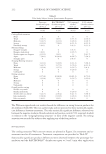
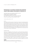
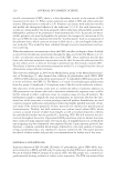
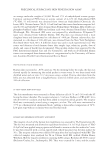
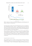
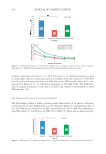
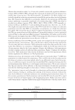
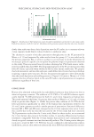
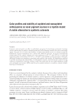
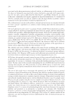
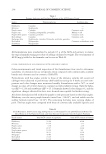
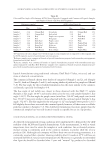
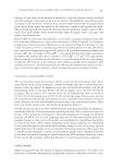
MEASUREMENT OF ADVANCED GLYCATION END-PRODUCTS 197 The default design of the instrument is to be used on a bench top for ease of measuring the volar forearm. To measure the facial skin, we obtained the instrument manufacturer’s technical approval and tilted the measuring surface by a 65° angle, thus allowing the left cheek of a subject to comfortably engage the measuring window. Figure 1 shows the con- fi guration of the new installation together with a picture of the traditional position. FACIAL SKIN COLOR AND WRINKLE MEASUREMENTS Facial skin color and wrinkle measurements were performed by using image analysis means. VISIA-CR® (Canfi eld Scientifi c, Parsippany, NJ) was used to capture facial images under fi ve different lighting conditions (standard, fl at, UV, cross polarized, and parallel polarized). Amway exclusive Facial Analysis Computer Evaluation System was used to objectively measure skin color parameters such as individual typology angle (ITA°) and facial wrinkles. The wrinkle measurement was from the frontal image of whole face, and the ITA° was measured from the cheek area (37). Figure 2 shows a sample output of wrinkle analysis result (A) and a region of interest on a facial image for skin color measurement (B). SUBJECTS A total of 239 healthy Caucasian volunteers, Fitzpatrick Skin Type II and III, aged 21- to 65-year old with 151 females (63.2%, mean age = 43.1) and 88 males (36.8%, mean age = 41.4) participated in the study. All subjects were provided with verbal and written descriptions regarding the intent of the study and each signed an informed consent form consistent with the requirements in the Code of Federal Regulations Title 21 (21 CFR) Fig ure 1. Set up of AGE Reader for skin AGE measurement. (A) For the cheek. (B) For the volar forearm.
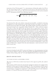
Purchased for the exclusive use of nofirst nolast (unknown)
From: SCC Media Library & Resource Center (library.scconline.org)