JOURNAL OF COSMETIC SCIENCE 222 were reported as μl 3 H2O/cm2. Samples with water permeation greater than 2.0 μl 3 H2O/ cm2 were discarded. The remaining cells were ranked in order of increasing water perme- ability to facilitate the random controlled block experimental design (8). The receptor exchange procedure was repeated, and the cells were allowed to wash out overnight. A fi nal exchange was performed in the morning before dosing. Porcine skin integrity was assessed visually to ensure the absence of large hair follicles. SURFACTANT PENETRATION PROTOCOL—PORCINE SKIN 14 C-SDS solutions (50 mM SLS + 6.7 μCi/ml 14 C-SDS) in DI water, with and without 2% w/w of added polymer, were prepared and shaken to ensure homogeneity. The SLS con- centration corresponds to 1.44% w/v, about 10-fold lower than typical anionic surfactant concentrations in a shampoo or shower gel. This is a commonly accepted dilution factor for consumer exposures. The test concentration was furthermore about 16-fold higher than the apparent CMC for the SLS sample, so most of the SLS in these formulations existed in either micellar or polymer-bound micellar form. A 150 μl aliquot (10 μCi) of the surfac- tant solution was pipetted onto each skin membrane. Skin from one donor was exposed to the surfactant solution for 10 min (n = 6/treatment). After the surfactant exposure, the dose solution was removed using a transfer pipet. The surface of the skin was rinsed three times with 0.5 ml of tap water for 10 s, and the rinses were collected and pooled. The receptor solution was collected, and each skin sample was wiped two times with Whatman fi lter paper (GE Healthcare Life Sciences, Pittsburgh, PA) soaked with PBS/Tween 20 and once with 70%/30% ethanol/water to remove unabsorbed (residual) product. Wipes were collected and pooled for mass bal- ance determination. After surface rinsing, the surfaces of the skins were dried, and 10 tape strips (D-Squame™) were collected. The tapes were placed directly into Ultima Gold XR cocktail to be analyzed individually. After tape stripping, the remaining epi- dermis was dissected from the dermis, and the skin sections were dissolved in 0.50–1.25 ml Soluene-350™ at 50°C overnight. Radioactivity in receptor collections, surface rinses, fi lter paper wipes, tape strips, and solubilized tissue sections was determined using LSC. Results were expressed as μg/cm2 14 C-SDS equivalents or % of applied radioactive dose. The arithmetic mean and standard error mean (SEM) were reported for each treatment. SURFACTANT PENETRATION PROTOCOL—HUMAN SKIN 14 C-SDS solutions (50 mM SLS + 6.7 μCi/ml 14 C-SDS) in DI water, with and without 2% w/w of an added polymer, were prepared and shaken to ensure homogeneity. A 150 μl aliquot (10 μCi) of the surfactant solution was pipetted onto each skin membrane, which were rank ordered in terms of permeability based on the 3 H2O prescreening results. The rankordering and subsequent randomization by treatment were key elements in maxi- mizing the sensitivity of the assay (8). Two sets of experiments were conducted. In Experiment 1, skin from three donors was exposed to the surfactant solution for 10 min (n = 4–6/donor). The total sample size was n = 14–15/treatment. In Experiment 2, skin from four donors was exposed to the surfac-

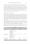
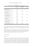
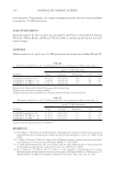
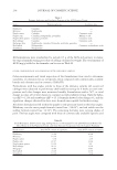
PRECLINICAL SURFACTANT SKIN PENETRATION ASSAY 223 tant solution for 2 min (n = 2–7/donor). The total sample size was n = 20–21/treatment. The treatment groups are summarized in Table I. The same method of collection was used as with porcine skin after the surfactant expo- sure. For Experiment 1, 4–10 tape strips were performed on each sample. The tapes were put directly into Ultima Gold XR cocktail and analyzed individually. The resid- ual epidermis and the dermis were also physically separated and dissolved overnight in Solvable™. For Experiment 2, neither tape stripping nor physical separation of the skin layers was conducted instead, the rinsed skin samples were removed from the dif- fusion cells and directly dissolved in 2 ml of Solvable™. This choice followed from the fact that Experiment 1 showed most of the residual radioactivity in the skin samples to be recovered in the fi rst three tape strips and only very low radioactivity levels in the lower skin layers. All samples were analyzed by LSC for 5 min or until 2% accuracy [2 relative standard deviations (SDs)] was reached. Results were expressed as microgram per square centimeter 14 C-SDS equivalents in the various samples, after background subtraction. The deposited dose was calculated as the total amount of 14 C-SDS in skin plus the receptor solution. HUMAN SKIN STATISTICAL ANALYSIS Outliers were detected using Dixon’s test on the full dataset following a logarithmic transformation of the individual sample values (8). Outlying results were rejected if they exceeded the 95% confi dence limit. The least squares mean and standard error for each skin donor were calculated these values were then averaged arithmetically over donors to obtain the fi nal mean and standard error. Results were expressed as microgram per square centimeter 14 C-SDS equivalents in the various samples, after background subtraction. Statistical comparisons between treat- ments were made via two-way ANOVA on the (log10)-transformed values, using skin donor and treatment as the blocking variables. There was a signifi cant statistical differ- ence between skin donors for both Experiments 1 and 2, yielding p 0.001 and p = 0.020, respectively. The SEM between donors ranged from 0.052 to 0.086 μg/cm2 for Experiment 1 and from 0.061 to 0.110 μg/cm2 for Experiment 2. Differences between treatments were highly signifi cant, with p 0.001 for Experiment 1 and p = 0.009 for Experiment 2. There were no signifi cant interactions between skin donor and treatment for either study. Therefore, differences attributed to skin donor did not depend on treat- ment and vice versa. Table I Do se Solutions and Sample Sizes for Human Skin 14 C-SDS Penetration Studies Treatments Sample size (n)a Expt. 1 10-min Expt. 2 2-min 50 mM SLS (control) 14 20 50 mM SLS + 2% PEO 15 21 50 mM SLS + 2% PVA 14 21 a Sum of replicate samples from 3 to 4 donors.
Purchased for the exclusive use of nofirst nolast (unknown)
From: SCC Media Library & Resource Center (library.scconline.org)