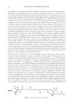
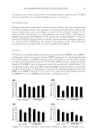
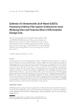
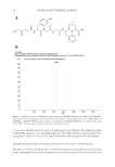
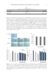
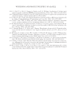
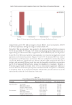
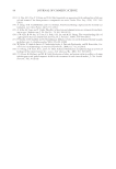
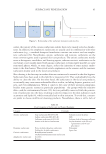
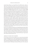
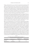
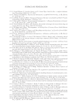
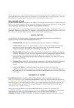
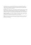
JOURNAL OF COSMETIC SCIENCE 58 RESULTS AND DISCUSSION CELL VIABILITY ASSAY Fu coxanthin has an exce llent antioxidant activity (29,30), and a recent study showed that fucoxanthin protects against Ultraviolet-B (UVB)-induced skin photoaging (23). In the present study, in vitro and in vivo experiments were conducted to evaluate the antiaging and antiwrinkle effects of a fucoxanthin concentrate prepared from P. tricornutum. Before effi cacy assessment, the cytotoxic effect of the concentrate was evaluated at various con- centrations to determine the appropriate concentration for treatment (Figure 2). The re- sults of the cytotoxicity experiment showed that PT-FX50 did not affect cell proliferation when it was used up to 25 μg/ml. However, it decreased cell number at concentrations of 50 and 100 μg/ml. Based on these results, subsequent experiments were conducted using the concentrate at d25 μg/ml. Fucoxanthin is reported to cause few adverse effects on normal cells at a low concentration (31–34). Liu et al. showed that at a concentration of 15 μg/ml, fucoxanthin inhibited the proliferation of glioma U87 and U251 cancer cell lines however, it was not cytotoxic up to 30 μg/ml to normal neurons (31). In another study, the half-maximum inhibitory concentration (IC50) values of pure fucoxanthin against the proliferation of human dermal fi broblasts, Human umbilical vein endothelial cell, and HEK293 cells after treatment for 48 h were found to be 32, 6.7, and 18.7 μM, respectively. The IC50 values of Undaria pinnatifi da extract containing 60.8% fucoxanthin against the three cell lines were 46.5, 6.7, and 33.9 μM, respectively (32). The 50% lethal dose of fucoxanthin in Institute of Cancer Research mice is reported to be 2,000 mg/kg body weight however, no obvious toxicity was observed after repeated oral administra- tion of pure fucoxanthin at 750 mg/kg to mice for 28 d (17,35). In addition, it has been demonstrated that fucoxanthin does not have a genotoxic effect on mouse bone marrow cells (36). Thus, although cell proliferation was inhibited by fucoxanthin at 50 μg/ml in Figure 2. Cytotoxicity of PT-FX50 against HDFN cells. Data are expressed as mean value ± standard deviation (n = 3).

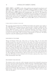
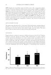
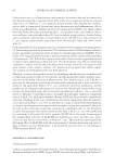
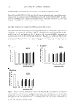
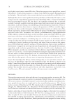
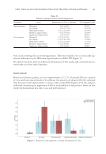
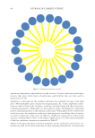
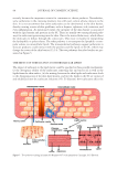
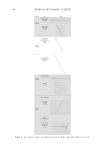
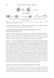
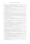
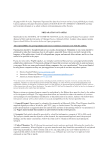
ANTIAGING POTENTIAL OF FUCOXANTHIN 59 the present study, based on the results of the aforementioned animal studies, PT-FX50 was not considered as an unsuitable ingredient for use in cosmetics. PROCOLLAGEN ASSAY Collagen is the main component of connective tissue. It plays a key role in maintaining the tensile strength of human skin. Collagen is produced as precursor forms called procol- lagens. Among them, type 1 procollagen accounts for 80% of dermal collagen (37). To assess the effect of fucoxanthin on collagenogenesis, the levels of type I procollagen in HDFN cells treated with PT-FX50 (3.1–25 μg/ml) were analyzed (Figure 3). It was ob- served that TGF-β (10 ng/ml) used as a positive control increased procollagen content in the cells by 29.0%. Furthermore, the amount of procollagen in the cells signifi cantly increased after the treatment with 12.5 and 25 μg/ml of fucoxanthin. MMP ASSAY UV rays increase wrinkle fo rmation by inducing the expression of MMPs, such as MMP-1 (collagenase), which cleaves type I collagen MMP-2 (gelatinase A), which degrades type IV and VII collagen and MMP-9 (gelatinase B), which degrades type IV and V collagen and other extracellular matrix proteins (38,39). Thus, MMPs are major markers of wrinkle formation. The MMP-1 level in the positive control cells treated with 10 μM RA was 47.2% less than that in the control cells, which means that MMP-1 levels decreased at a statistically signifi cant level (Figure 3). Furthermore, 25 μg/ml of fucoxanthin signifi cantly decreased MMP-1 and MMP-2 levels by 12.7% and 22.3%, respectively. Fucoxanthin decreased the MMP-9 level in the HDFN cells at all the concentrations tested. Figure 3. In vitro effi cacy of PT-FX50: (A) Procollagen, (B) MMP-1, (C) MMP-2, and (D) MMP-9 levels.
Purchased for the exclusive use of nofirst nolast (unknown)
From: SCC Media Library & Resource Center (library.scconline.org)