JOURNAL OF COSMETIC SCIENCE 68 controls. After incubation for 48 h, the absorbance was measured at 620 nm with a micro- plate reader (EZ read 400, Biochrom Ltd., Cambridge, UK). The results were transformed to a percentage of the controls. The IC50 measure was graphically obtained from the dose– response curves. CELL CULTURE AND CYTOTOXICI TY B16F10 melanoma cells and H aCaT keratinocyte cells that were purchased from the American Type Culture Collection (ATCC, Manassas, VA) were cultured in a 5% CO2 incubator at 37°C. The cells were cultured in DMEM supplemented with 10% FBS and 0.01% antibiotic-antimycotic solution (Invitrogen, Grand Island, NY) in a 5% CO2 incuba- tor at 37°C. The cytotoxic effect of 5-AL A and ALACELL was measured by the MTT assay (20). Briefl y, cells were seeded uniformly at 2.5 × 10⁴ cells/well densities in 96-well micro- plates. After 24 h, the media were replaced with 100 μL media containing fi nal concen- trations equivalent to 10, 20, and 40 μM in 5-ALA and ALACELL. The MTT assay was performed after 48 h of incubation with 5-ALA and ALACELL. The cytotoxicity was quantifi ed by measuring UV absorbance at 570 nm by a Tecan microplate reader (Mannedorf, Switzerland). The measured absorbance was standardized to the absorbance of nontreated control cells. All data represent the mean and standard deviation from at least three separate experiments and were compared using a student’s t-test. EFFECT OF ALACELL ON MELANIN FORM ATION The melanin formation was analyze d by a modifi cation of the method described by a previous study (21). B16F10 melanoma cells were seeded at a density of 3 × 10⁵ cells/well in 96-well microplates and cultivated by the method described earlier. To induce hyper- production of melanin, the cells were treated with α-MSH of 100 nM. Then, the concen- trations of 10, 20, and 40 μM of 5-ALA and ALACELL were added to the medium and further incubated for 48 h. To remove melanin excreted from the cells, the medium was removed, and the cells were washed twice with PBS, and then harvested by trypsin treat- ment. The harvested cells were pelleted, and the cell membrane was dissolved in Triton X-100 (Sigma-Adrich, St. Louis, MO). The purifi ed melanin was dissolved in 2 M NaOH for 30 min at 100°C. The absorbance was measured at 405 nm. The melanin content was compared with untreated control cells. EFFECT OF ALACELL ON INTRACELLULAR TY ROSINASE ACTIVITY Intracellular tyrosinase activity can rapidly oxidize L-tyrosine to L-3,4-dihydroxyphenyl- alanine (L-DOPA) and further convert it to dopaquinone, and then the activity of tyrosi- nase determines the amount of brown dopaquinone (22,23). In this study, B16F10 melanoma cells were seeded at a density of 3 × 10⁵ cells/well in 96-well microplates, and the culture medium was discarded after 24 h. Five-ALA and ALACELL were treated with 10, 20, and 40 μM. After 48 h, the cells were washed twice with PBS. B16F10 melanoma cells were lysed in 1% Triton X-100, 20 mM sodium phosphate (pH 6.8), and 1 mM

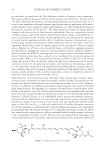
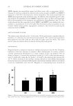
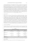
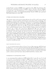
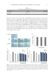
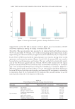
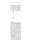
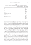
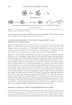
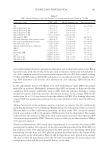
WHITENING AND PROTECTIVE EFFECT OF ALACELL 69 PMSF, and centrifuged at 14,000 rpm for 15 min. The Bradford assay with bovine serum albumin as the protein standard was conducted to measure the protein content of the supernatant. Intracellular tyrosinase activity was measured in a reaction mixture (1 mL) containing 50 mM phosphate buffer (pH 6.8), 2 mM L-DOPA, and 300 μg supernatant proteins. After incubation at 37°C for 15 min, the absorbance was measured at 475 nm by a microplate reader. Tyrosinase activity (%) = [(A-B)/C] × 100% A: sample absor- bance volume, B: blank absorbance volume, and C: control absorbance volume. EFFECT OF ALACELL ON UVB-IRRADIATED HaCaT CELL S UVB irradiation was performed by UVM-225D Mine ralight UV display lamps (UVP, Phoenix, AZ) generating UVB light in the range of 290–320 nm with a maximum emis- sion wavelength of 302 nm. UV doses were measured using a HD2102-2 UV meter (Delta OHM, Padova, Italy). HaCaT keratinocyte cells were seeded on six-well plates at 7 × 10⁴ cells/well and incubated in a 5% CO2 incubator at 37°C. Five-ALA and ALACELL were treated in cultured cells at concentrations of 20, 40, 80, and 100 μM, and after 1 h, the cells were irradiated with UVB at 40, 80, and 120 mJ/cm2. To prevent UV quenching, before irradiation, the cell culture medium was replaced by the same volume of PBS after two washing steps with PBS. After UVB irradiation, cells were fed with fresh growth medium and incubated. After 24 h, the cell viability was measured by the MTT assay described earlier. STATISTICAL ANALYSIS Results are expressed as mean s ± standard deviatio n. Data were analyzed using analysis of variance and Duncan’s multiple range tests. Signifi cance was indicated at *p 0.05, **p 0.01, and ***p 0.001. RESULTS CONFIRMATION OF ALACELL STRUCTURE BY MALDI-T OF MS AL ACELL was synthesized by combining the phytochemic al agent 5-ALA with Y-G-G- F-L peptide to support physicochemical activity. MALDI-TOF mass spectrometry was performed to identify the molecular weight and chemical structure of ALACELL (Figure 1A). The chemical formula of ALACELL is C33H44N6O9, and its molecular weight is 668.32 (Figure 1B). GROWTH INHIBITORY EFFECT OF ALACELL AGAINST C. ACNES To investigate the antimicrobial activities of 5-ALA and ALACELL, we determined the minimum concentration that produces 50% inhibition of bacterial growth (IC50) by the broth dilution assay. We tested different concentrations for each sample against C. acnes, S. aureus, B. cereus, E. coli, and Y. enterocolitica. 5-ALA and ALACELL against C. acnes blocked growth dose dependently, respectively (Table I). The IC50 of 5-ALA and ALACELL against
Purchased for the exclusive use of nofirst nolast (unknown)
From: SCC Media Library & Resource Center (library.scconline.org)