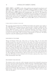
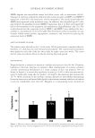
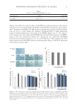
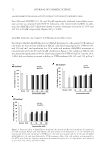
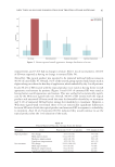
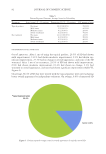
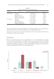
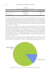
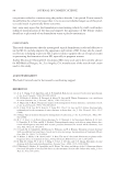
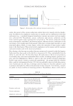
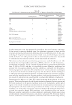
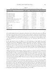
JOURNAL OF COSMETIC SCIENCE 84 Al l of these ingredients used in the product we evaluated have been individually shown to improve SD in prior literature and, now, when collectively used in our topical formula- tion, SD improved signifi cantly within 1 mo of use. We are unable to discern if there is a specifi c ingredient that may play a dominant role in the formulation, but it is most likely the improvement we saw is because of the synergistic effect of all the ingredients working along the surface of the skin. Th ere is evidence to support the use of silicone gel in treating SD. A recent study found that SD treated with silicone over a placebo cream had a statistically increased collagen and reduction in pigmentation (13). In addition, 2 recent studies demonstrated that silicone-based products improves SD in pregnant women (9,21). SD have histological similarities to scars, and silicone, which is used as the foundational matrix of our product, has a long track record as a scar-reducing treatment (24). In clinical trials, silicone has demonstrated effectiveness in preventing hypertrophic or keloid scarring in patients with newly healed wounds (25). I n addition, growth factors play a role in the progression of SD. A recent study indicated the positive effects of using growth factors to manage SD (12). Moreover, from a histo- logic standpoint, SD formation is similar to wound-healing processes found in scars Table VII Evaluator Average Scores for Overall Appearance Side Proportion (%) 95% margin of error Topical product better 19/22 (86.4%) ±14.3% Same 3/22 (13.6%) ±14.3% No treatment better 0/22 (0.0%) ±0.0% Figure 5. Evaluator average sc o res for overall appearance of topically treated SD.

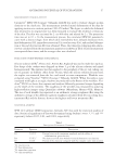
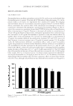
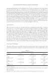
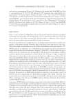
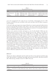
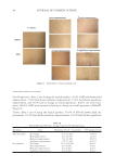
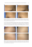
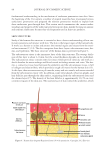
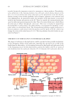
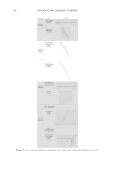
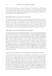
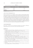
NEW TOPICAL SILICONE FORMULATION FOR TREATING STRIAE DISTENSAE 85 (4,26–28). The initial infl ammatory, pruritic plaques evolve into stable, atrophic hypopig- mented streaks that can be considered fi nal scar tissue following the prior infl ammatory changes. Anti-infl ammatory and pro-infl ammatory growth factors play a role in the tran- sition of SD, and many believe that a disruption of the TGF-β growth factor messenger Figure 6. Three-dimensional pho t ographic comparison of abdominal SD before and after 1-mo treatment with the topical product on one side (A) and no treatment on the other side (B). There is signifi cant improve- ment in the overall appearance of SD on the side treated with the topical product (C). By contrast, the side left untreated (D) does not show improvement. Figure 7. Three-dimensional photo graphic comparison of thigh SD before and after 1-mo treatment with the topical product on one side (A) and no treatment on the other side (B). There is moderate improvement in the overall appearance of SD on the side treated with the topical product (C). By contrast, the side left untreated (D) does not show improvement.
Purchased for the exclusive use of nofirst nolast (unknown)
From: SCC Media Library & Resource Center (library.scconline.org)