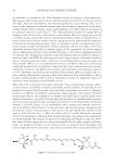
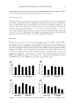
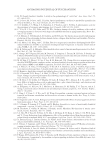
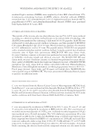
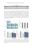
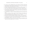
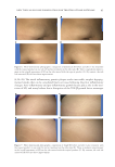
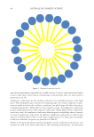
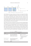
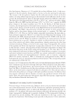
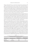
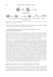
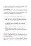
JOURNAL OF COSMETIC SCIENCE 54 an imbalance in metabolism (6). The imbalance results in changes in skin appearance. Skin aging is affected by genetic factors and the external environment (1). Factors such as UV light, hormonal abnormality, environmental pollutants, and smoking cause an in- crease in the expression of dermal enzymes that degrade major components of the extra- cellular matrix, such as collagen, elastin, and hyaluronic acid. This results in alteration of the physical structure of the skin (7). The most prominent features of aging-induced changes in the skin are loss of skin elasticity and wrinkles. Reactive oxygen species such as singlet oxygen, superoxide radical, and hydroxyl radical, which are generated by in- trinsic and extrinsic factors, mediate the skin aging process by promoting the expression of several matrix-degrading enzymes (8,9). A signifi cant amount of superoxide is pro- duced during normal mitochondrial cellular respiration (10,11). UV light is the most important external factor that accelerates aging via the generation of reactive oxygen species. Exposure to UV rays can be avoided by using a cover cloth or applying sunscreen on exposed skin. Endogenous reductases and antioxidants prevent oxidative damage to proteins, lipids, and DNA by reducing toxic active oxygen species to harmless chemical species. Several studies have been carried out to investigate how to prevent aging and treat wrinkles. Weiss et al. (12) reported that retinoic acid (RA) is effective in alleviating rough and wrinkled skin. In addition, other clinically active substances such as retinol, vitamin C, vitamin E, and adenosine are used as raw materials in formulating cosmetics (13–15). However, the need for raw materials with excellent effi cacy is high in the cos- metic industry. Research is ongoing to fi nd such substances from natural sources this is because natural products with excellent antioxidant activity are important sources of substances with antiaging and antiwrinkle effects. Mor e than 600 carotenoids have been identifi ed. They include beta-carotene, alpha- carotene, lutein, zeaxanthin, lycopene, astaxanthin, and fucoxanthin. Carotenoids are very benefi cial to human health because they have high antioxidant activity (16). However, because it is diffi cult to synthesize carotenoids in large amounts, they must be obtained from natural sources. Fucoxanthin is a common carotenoid that is mostly found in the marine environment, especially in brown algae and diatoms. Fucoxanthin makes up more than 10% of the carotenoids that are produced in nature (17,18). Fucoxanthin (molecular formula, C42H58O6 Figure 1) is a xanthophyll with a unique polyisoprenic structure, which includes an allene bond a 5,6-monoepoxide and a ketone group conjugated with the polyenic system. It has a brown or olive-green color and signifi cantly absorbs visible light at 400–500 nm (λmax = 449 nm) within the UV–visible absorption spectrum (19,20). Fucoxanthin is commonly found in photosynthetic microalgae and is responsible for absorbing light. Microalgae are exposed to strong light and oxygen conditions there- fore, many reactive oxygen species, such as superoxide radical, singlet oxygen, and nitric oxide, are produced in them (21,22). Extracts that are prepared from microalgae and contain high amounts of carotenoids, especially fucoxanthin, show strong antioxidant and Figure 1. Chemical structure of fucoxanthin.

ANTIAGING POTENTIAL OF FUCOXANTHIN 55 anti-infl ammatory activities (21). Fucoxanthin has various physiological activities such as anticancer, anti-obesity, antiangiogenic, and neuroprotective activities (18,23–26). The aim of this study was to investigate the antiaging effect of fucoxanthin concentrate derived from Phaeodactylum tricornutum and evaluate its potential use in wrinkle care cosmetics. MATER IAL AND METHODS MATER IALS Phaeo dactylum tricornutum concentrate containing ≥50% fucoxanthin (PT-FX50) was pro- vided by Systems Biotechnology Research Center of the Korea Institute of Science and Technology (Gangneung, Korea) (27). High-performance liquid chromatography (HPLC)–mass spectrometry and HPLC–photodiode array detection (PDA) were used to assay the amount of fucoxanthin in P. tricornutum concentrate powder qualitatively and quantitatively. The fucoxanthin content in the powder was found to be 51.5%. In addition, P. tricornutum concentrate was diluted to 2% with caprylic/capric triglycerides (PT-FX01) and used as the fucoxanthin raw material to prepare a wrinkle care cream. The concentra- tion of fucoxanthin in PT-FX01 was determined to be 1.01% by HPLC-PDA. CELL V IABILITY ASSAY Cell v iability was measured using EZ-Cytox enhanced cell viability kit (Daeil Lab Ser- vice, Seoul, Korea) according to the manufacturer’s instructions. In brief, human dermal fi broblast, neonatal (HDFN) cells were cultured in Dulbecco’s modifi ed Eagle medium (DMEM) containing 10% fetal bovine serum, 1% penicillin/streptomycin, and 4 mM L-glutamine at 37°C in a humidifi ed atmosphere of 5% CO2. The cells were seeded at a density of 1 × 10⁴ cells/well in a 96-well plate and cultured for 24 h. PT-FX50 was dis- solved in dimethyl sulfoxide to prepare a 10% stock solution, which was serially diluted with DMEM and added to the culture medium to treat with 3.125, 6.25, 12.5, 25, 50, and 100 ppm of PT-FX50. Cell viability was determined 24 h later. The EZ-Cytox reac- tion mixture (Ez-3000, Daeil Lab Service) was diluted 10-fold by mixing it with the medium. Next, 100 μL of the diluted reaction mixture was added to each well. After 1 h of incubation, cell viability was determined by measuring absorbance at 450 nm and expressing it as a percentage relative to the viability of untreated cells. PROCOLLAG EN, MATRIX METALLOPROTEINASE (MMP)-1, MMP-2, AND MMP-9 ASSAYS HDFN cell s were seeded at a density of 1 × 10⁴ cells/well in a 96-well plate and cultured for 24 h. The medium was removed and kept for 24 h in a starvation state. Next, the stock PT-FX50 solution was diluted with DMEM to different concentrations, after which the cells were treated and cultured for 24 h. The culture medium was harvested, and the amount of procollagen in it was measured using procollagen type I c-peptide EIA kit (Mk1010 Takara, Shiga, Japan). 10 ng/ml of transforming growth factor (TGF)-β was used as a positive control for procollagen synthesis assay. In addition, the amounts of
Purchased for the exclusive use of nofirst nolast (unknown)
From: SCC Media Library & Resource Center (library.scconline.org)