JOURNAL OF COSMETIC SCIENCE 338 ( 42) K. A. Jay, D. L. Smith, and E. H. Blackburn, Early loss of telomerase action in yeast creates a depen- dence on the DNA damage response adaptor proteins, Mol. Cell Biol., 3, 1908–1919 (2016). ( 43) E. M. Buckingham and A. J. Klingelhutz, The role of telomeres in the ageing of human skin, Exp. Dermatol., 20, 297–302 (2011). ( 44) S. S. Jose, K. Bendickova, T. Kepak, Z. Krenova, and J. Fric, Chronic infl ammation in immune aging: role of pattern recognition receptor crosstalk with the telomere complex? Front. Immunol., 8, 1078– 1078 (2017). ( 45) E. Goyarts, N. Muizzuddin, D. Maes, and P. U. Giacomoni, Morphological changes associated with aging. Ann. N. Y. Acad. Sci., 1119, 32–39 (2007). ( 46) A. Slusher, T. Zúñiga, and E. Acevedo, Infl amm-aging is associated with lower Plasma PTX3 concen- trations and an impaired capacity of PBMCs to express hTERT following LPS stimulation, Mediat. Infl amm., 2019, 1–13 (2019). (4 7 ) A. Kraag Ziegler, A. Damgaard, A. Mackey, P. Schjerling, S. Magnusson, A. Olesen, M. Kjaer, and C. Scheele, An anti-infl ammatory phenotype in visceral adipose tissue of old lean mice, augmented by exercise, Sci. Rep., 9, 12069 (2019). (4 8 ) F. Mangiola, A. Nicoletti, A. Gasbarrini, and F. R. Ponziani, Gut microbiota and aging, Eur. Rev. Med. Pharmacol. Sci., 22, 7404–7413 (2018). (4 9 ) D. Mariat, O. Firmesse, F. Levenez, V. Guimaraes, H. Sokol, J. Dore, G. Corthier, and J. P. Furet, The Firmicutes/Bacteroidetes ratio of the human microbiota changes with age, BMC Microbiol., 9, 123 (2009). (5 0 ) N. Thevaranjan, A. Puchta, C. Schulz, A. Naidoo, J. C. Szamosi, C. P. Verschoor, D. Loukov, L. P. Schenck, J. Jury, K. P. Foley, J. D. Schertzer, M. J. Larche, D. J. Davidson, E. F. Verdu, M. G. Surette, and D. M. E. Bowdish, Age-associated microbial dysbiosis promotes intestinal permeability, systemic infl ammation, and macrophage dysfunction, Cell. Host. Microbe., 23, 570 (2018). (5 1 ) N. Shibagaki, W. Suda, C. Clavaud, P. Bastien, L. Takayasu, E. Iioka, R. Kurokawa, N. Yamashita, Y. Hattori, C. Shindo, L. Breton, and M. Hattori, Aging-related changes in the diversity of women’s skin microbiomes associated with oral bacteria, Sci. Rep., 7, 10567 (2017). (5 2 ) K. Neha, M. R. Haider, A. Pathak, and M. S. Yar, Medicinal prospects of antioxidants: a review, Eur. J. Med. Chem., 178, 687–704 (2019). (5 3 ) O. Ighodaro and O. Akinloye, First line defence antioxidants-superoxide dismutase (SOD), catalase (CAT) and glutathione peroxidase (GPX): their fundamental role in the entire antioxidant defence grid, Alexandria J. Med., 54, 287–293 (2018). (5 4 ) H. Masaki, Role of antioxidants in the skin: anti-aging effects, J. Dermatol. Sci., 58, 85–90 (2010). (5 5 ) H. Lee, N. Park, and Y. Hwang, A new dimension of the digital divide: exploring the relationship between broadband connection, smartphone use and communication competence, Telematics Inform., 32, 45–56 (2015). (5 6 ) L. Vollono, M. Falconi, R. Gaziano, F. Iacovelli, E. Dika, C. Terracciano, L. Bianchi, and E. Campione, Potential of curcumin in skin disorders, Nutrients, 11, 2169 (2019). (5 7 ) B. Sommerfeld, Randomised, placebo-controlled, double-blind, split-face study on the clinical effi - cacy of Tricutan on skin fi rmness, Phytomedicine, 14, 711–715 (2007). (5 8 ) R. M. Martinez, F. A. Pinho-Ribeiro, V. S. Steffen, T. C. Silva, C. V. Caviglione, C. Bottura, M. J. Fonseca, F. T. Vicentini, J. A. Vignoli, and M. M. Baracat, Topical formulation containing naringenin: effi cacy against ultraviolet B irradiation-induced skin infl ammation and oxidative stress in mice, PloS One, 11, e0146296 (2016). (5 9 ) W. N. Yap, Tocotrienol-rich fraction attenuates UV-induced infl ammaging: a bench to bedside study, J. Cosmet. Dermatol., 17, 555–565 (2018). (60) E . J. Kovacs, Aging, traumatic injury, and estrogen treatmen, Exp. Gerontol., 40, 549–555 (2005). (61) P . Lencel and D. Magne, Infl ammaging: the driving force in osteoporosis? Med. Hypotheses, 76, 317– 321 (2011). (62) M . P. Brincat, Hormone replacement therapy and the skin, Maturitas, 35, 107–117 (2000). (63) M . Brincat, A. W. Yuen, J. W. Studd, J. Montgomery, A. L. Magos, and M. Savvas, Response of skin thickness and metacarpal index to estradiol therapy in postmenopausal women, Obstet. Gynecol., 70, 538–541 (1987). (64) M . B. Stout, F. J. Steyn, M. J. Jurczak, J. G. Camporez, Y. Zhu, J. R. Hawse, D. Jurk, A. K. Palmer, M. Xu, T. Pirtskhalava, G. L. Evans, R. de Souza Santos, A. P. Frank, T. A. White, D. G. Monroe, R. J. Singh, G. Casaclang-Verzosa, J. D. Miller, D. J. Clegg, N. K. LeBrasseur, T. von Zglinicki, G. I. Shulman, T. Tchkonia, and J. L. Kirkland, 17alpha-Estradiol alleviates age-related metabolic and infl ammatory dys- function in male mice without inducing feminization, J. Gerontol. A Biol. Sci. Med. Sci., 72, 3–15 (2017).

SKIN-AGING AND INFLAMMAGING TREATMENT 339 (65) L . A. Brinton, D. R. Brogan, R. J. Coates, C. A. Swanson, N. Potischman, and J. L. Stanford, Breast cancer risk among women under 55 years of age by joint effects of usage of oral contraceptives and hormone replacement therapy, Menopause, 25, 1195–1200 (2018). (66) F . Laliberté, K. Dea, M. S. Duh, K. H. Kahler, M. Rolli, and P. Lefebvre, Does the route of admin- istration for estrogen hormone therapy impact the risk of venous thromboembolism? Estradiol transdermal system versus oral estrogen-only hormone therapy, Menopause, 25, 1297–1305 (2018). (67) Z . Fazeli, A. Abedindo, M. D. Omrani, and S. M. H. Ghaderian, Mesenchymal stem cells (MSCs) therapy for recovery of fertility: a systematic review, Stem Cell Rev. Rep., 14, 1–12 (2018). (68) J . Doles, M. Storer, L. Cozzuto, G. Roma, and W. M. Keyes, Age-associated infl ammation inhibits epidermal stem cell function, Genes Dev., 26, 2144–2153 (2012). (69) A . Mojallal, C. Lequeux, C. Shipkov, P. Breton, J. L Foyatier, F. Braye, and O. Damour, Improvement of skin quality after fat grafting: clinical observation and an animal study, Plast. Reconstr. Surg., 124, 765–774 (2009). (70) S . Zhang, Z. Dong, Z. Peng, and F. Lu, Anti-aging effect of adipose-derived stem cells in a mouse model of skin aging induced by D-galactose, PloS One, 9, e97573 (2014). (71) H . J. Kim and J.-S. Park, Usage of human mesenchymal stem cells in cell-based therapy: advantages and disadvantages, Dev. Reprod., 21, 1–10 (2017). (72) T . Quan and G. J. Fisher, Role of age-associated alterations of the dermal extracellular matrix micro- environment in human skin aging: a mini-review, Gerontology, 61, 427–434 (2015). (73) L . F. Lau, CCN1/CYR61: the very model of a modern matricellular protein, Cell Mol. Life Sci., 68, 3149 (2011). (74) R . Kafi , H. S. R. Kwak, W. E. Schumacher, S. Cho, V. N. Hanft, T. A. Hamilton, A. L. King, J. D. Neal, J. Varani, and G. J. Fisher, Improvement of naturally aged skin with vitamin a (retinol), Arch. Dermatol., 143, 606–612 (2007). (75) K . Rehman and M. H. Zulfakar, Recent advances in gel technologies for topical and transdermal drug delivery, Drug Dev. Ind. Pharm., 40, 433–440 (2014). (76) A . -R. Denet, R. Vanbever, and V. Préat, Skin electroporation for transdermal and topical delivery, Adv. Drug Deliv. Rev., 56, 659–674 (2004). (77) G. Sahu, S. Sahu, H. Sharma, and A. K. Jha, A review of current and novel trends for anti-ageing formulation, Int. J. Pharm. Chem. Biol. Sci., 4, 118–125 (2014). (78) N. A. Patel, N. J. Patel, and R. P. Patel, Formulation and evaluation of curcumin gel for topical ap- plication, Pharm. Dev. Technol., 14, 83–92 (2009). (79) W. R. Pfi ster and D. S. Hsieh, Permeation enhancers compatible with transdermal drug delivery sys- tems. Part I: selection and formulation considerations, Med. Device Technol., 1, 48–55 (1990). (80) I. Rodríguez Cruz, C. Domínguez-Delgado, J. Escobar-Chávez, M. López-Cervantes, and R. Díaz- Torres, Physical Penetration Enhancers: an Overview (Bentham Science Publishers, Sharja, United Arab Emirates, 2016), pp. 3–34. (81) Y. Chen, M. Wang, and L. ang, Biomaterials as novel penetration enhancers for transdermal and der- mal drug delivery systems, Drug Deliv., 20, 199–209 (2013). (82) Y.-W. H o u, M.-H. Chan, H.-R. Hsu, B. R. Liu, C.-P. Chen, H.-H. Chen, and H.-J. Lee, Transdermal delivery of proteins mediated by non-covalently associated arginine-rich intracellular delivery pep- tides, Exp. Dermatol., 16, 999–1006 (2007). (83) Y. Z. L i , Y. S. Quan, L. Zang, M. N. Jin, F. Kamiyama, H. Katsumi, A. Yamamoto, and S. Tsutsumi, Transdermal delivery of insulin using trypsin as a biochemical enhancer, Biol. Pharm. Bull., 31, 1574– 1579 (2008). (84) S. A. Nasrollahi, S. Fouladdel, C. Taghibiglou, E. Azizi, and E. S. Farboud, A peptide carrier for the delivery of elastin into fi broblast cells, Int. J. Dermatol., 51, 923–929 (2012). (85) Ge X., Wei M., He S., and Yuan W.-E., Advances of non-ionic surfactant vesicles (niosomes) and their application in drug delivery, Pharmaceutics, 11, 55 (2019). (86) B. Maherani, E. Arab-Tehrany, M. R Mozafari, C. Gaiani, and M. Linder, Liposomes: a review of manufacturing techniques and targeting strategies, Curr. Nanosci., 7, 436–452 (2011). (87) A. Laouini, C. Jaafar-Maalej, I. Limayem-Blouza, S. Sfar, C. Charcosset, and H. Fessi, Preparation, charac- terization and applications of liposomes: state of the art, J. Colloid Sci. Biotechnol., 1, 147–168 (2012). (88) L. Tavano, “Liposomal gels in enhancing skin delivery of drugs,” in Percutaneous Penetration Enhanc- ers Chemical Methods in Penetration Enhancement: Drug Manipulation Strategies and Vehicle Ef- fects, N. Dragicevic and H. I. Maibach. Eds. (Springer Berlin Heidelberg: Berlin and Heidelberg, 2015), pp. 329–341.
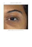
Purchased for the exclusive use of nofirst nolast (unknown)
From: SCC Media Library & Resource Center (library.scconline.org)