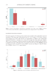
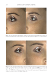
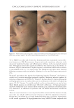
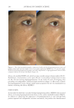
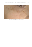
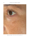
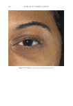
SKIN-AGING AND INFLAMMAGING TREATMENT 323 INTRINSIC AGING Intrinsic aging represents the inevitable and genetically determined aging of all tissues (17). It is shaped by endogenous physiological determinants, including gender, ethnicity, anatomical differences, and hormonal fl uctuations (18). Clinical signs of intrinsic skin aging include xerosis, fi ne lines, decreased elasticity, and subepidermal atrophy (17). These changes are based on reduced cellular proliferative capacity and genetic abnormalities (19). In addition, the dermis of older skin is characterized by fewer mast cells, fi broblasts, elastic fi bers, and lower amounts of collagen compared with younger skin (20). Moreover, signs of intrinsic skin aging not only include declines in fi brous extracellular matrix components, such as elastin, fi brillin, and collagens but also degeneration of oligosaccharides, which infl uence the skin’s capacity to preserve bound water (21). Although these structural changes are natural aspects of skin aging, environmental and individual factors, such as UV exposure and diet, can dramatically infl uence the rate of skin aging (22). EXTRINSIC AGING Extrinsic aging is referred to as photoaging, as it is shaped by environmental causes, es- pecially UV exposure (22). In fact, UV radiation exposure is considered the key determi- nant of extrinsic skin aging and attributable to 80% of facial skin aging (2). Whereas intrinsic aging causes epidermal thinning, photoaging is characterized by epidermal thickening based on impaired keratinocyte differentiation in the epidermal layer and basal cells (23). In addition, keratinocyte proliferation is impaired in both stratum cor- neum (SC) and basal cells (24). Furthermore, accelerated skin aging is associated with increased levels of MMPs (an enzyme family responsible for the decay of collagen and extracellular matrix proteins) (25). Although elastic fi ber degradation is a characteristic feature associated with aging, pho- toaging exhibits enormous accumulation of dystrophic elastin in the dermis known as solar elastosis (26). Elastin degradation in photoaging could be due to MMP activation, specifi cally human macrophage metalloelastase secreted by keratinocytes, fi broblasts, and infl ammatory cells (27). Mora Huertasa et al. (28) investigated the molecular changes in elastin associated with normal aging and photoaging. The study revealed that the elastin cleavage pattern is different in both types and is more pronounced in photoaging. More- over, the N-terminal of tropoelastin becomes more susceptible to enzymatic degradation due to photoaging (28). Moreover, air pollution has detrimental effects on skin. Air pollutants (ozone, volatile organic compounds, oxides, and others) alter skin homeostasis and induce aging and other infl ammatory diseases. This infl uence could be attributed to different mechanisms includ- ing free radical production, infl ammatory mediator release, and skin barrier damage (29). MOLECULAR PATHWAYS AND PROCESSES OF SKIN INFLAMMAGING It is important to identify molecular mechanisms, which are likely complementary and interconnected, that control skin aging to fi nd benefi cial approaches for prevention and treatment. Infl ammaging plays a crucial role in age-related diseases, such as osteoporosis,

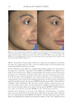
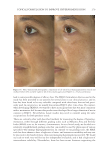
JOURNAL OF COSMETIC SCIENCE 324 Alzheimer’s disease, diabetes type II, and skin aging. Chronic exposure to intrinsic and extrinsic factors by itself generates the release of infl ammatory mediators, causing infl am- mation as well as dermal and extracellular matrix damage (2). The next section highlights the main pathways of skin infl ammaging, considering DNA damage and oxidative stress as key players in the process. OXIDATIVE STRESS Oxidative stress, which occurs because of a mismatch between ROS production and the cell’s ability to detoxify these species (30), is a key contributor to skin aging (31). ROS are generated by-products of oxygen metabolisms in every cell from different sources, including mitochondria, peroxisomal activity, oxidase activity, and endoplasmic reticu- lum (ER) (32). ROS at normal levels have benefi cial functions for the body, including cellular structure synthesis, fi ghting pathogens, and numerous signaling pathways (30). However, if ROS levels increase, oxidative stress occurs, harming cellular structures and immune responses and accelerating skin aging (30). Under normal conditions, receptor protein tyrosine phosphatases (RPTPs) inhibit recep- tor tyrosine kinase (RTK) activity on the cell surface through dephosphorylation (33). However, under oxidative stress, ROS bind to cysteine of RPTPs, inhibiting its activity and increasing phosphorylated RTKs levels. Consequently, numerous downstream signal- ing pathways are triggered, such as initiation of mitogen-activated protein kinase, tran- scription factor activator protein-1 (AP-1), and nuclear factor-κB (NF-κB) (34). The process inhibits collagen production and increases MMP gene transcription (2,35). Col- lagen degradation by MMPs leads to a build-up of fragmented and disorganized collagen fi brils, which downregulate new collagen synthesis (25). MMP-1, MMP-3, and MMP-9 collectively account for most of the type I and III dermal collagen degradation (36). In addition, reduced collagen content is attributable to AP-1’s suppression of type I and III procollagen gene expression in the dermis (37). D ifferent studies have reported a strong correlation between oxidative stress and infl am- mation, as continued exposure to ROS induces cell damage and, subsequently, a proin- fl ammatory signaling response. Oxidative damage to cells triggers TNF-α release, which in turn binds to cell surface receptors, activating the NF-κB infl ammasome. NF-κB in- fl ammasome generation results in an overproduction of other proinfl ammatory cytokines, which can be detrimental to health (38,39). In addition, UV radiation activates infl am- matory mediators, such as neutrophils, to remove damaged cells. Moreover, macrophages infi ltrate the exposed area, release ROS, and degrade the extracellular matrix (2,35). This is accompanied by fi broblast deterioration and an inability to repair the extracellular matrix, leading to skin aging (40). Therefore, NF-κB infl ammasome is considered the major etiology of infl ammaging. MICRO INFLAMMATORY THEORY The m icroinfl ammatory theory describes skin aging as a number of events in a repeated cycle that occurs because of cell exposure to intrinsic or extrinsic factors. (i) Damaged cells secrete proinfl ammatory signals such as prostaglandins and leukotrienes. (ii). These
Purchased for the exclusive use of nofirst nolast (unknown)
From: SCC Media Library & Resource Center (library.scconline.org)