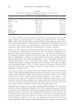
JOURNAL OF COSMETIC SCIENCE 368 Ceramides can be used as a moisturizer to treat dry skin. Many kinds of ceramides can be used synergistically to improve skin barrier function (4). Ceramides can also reduce the severity of skin diseases, reduce pain, improve the effi cacy of other drugs, reduce adverse reactions to drugs, accelerate the recovery of diseases, and improve quality of life (5). Ceramides have been used in topical formulations of AD (6). Ceramides can also reduce the reactivity to environmental pollution and skin sensitivity (7). Ceramide in the blood is used as a biomarker for predicting cardiovascular diseases (8) and pulmonary cystic fi brosis (9) and other diseases. Increase of ceramide contents in the blood may also be related to the risk of Alzheimer’s disease and can predict cognitive impairment (10). Therefore, determi- nation of the ceramide content qualitatively and quantitatively is very important for assess- ing skin condition and occurrence and development of various diseases. Busman (11) measured intracellular ceramide species using high-performance liquid chroma- tography (HPLC) coupled to atmospheric pressure-ionization mass spectrometry (MS). Zhixin (12) extracted human epidermis SC using tape stripping, and then quantifi ed ceramides using normal-phase liquid chromatography combined with dynamic multi-reaction monitoring MS. Twelve ceramide subclasses were found, including CER[NDS], CER[NS], CER[NP], CER[NH], CER[ADS], CER[AS], CER[AP], CER[AH], CER[EODS], CER[EOS], CER[EOP], and CER[EOH]. Here, we identifi ed the ceramide kinds and measured its content in skin and blood samples using HPLC and high-resolution MS and then analyzed them. The difference in ceramide in different tissues has not been detected by using precise methods. The high-throughput mass spectrometer has detected ceramide, and the detection result has high accuracy and the detection speed is very fast. MATERIALS AND METHODS SUBJECTS Subject inclusion criteria: healthy Chinese volunteers (18–65 y old, no gender requirements), no history of skin disease, no skin care moisturizers or other topical preparations used within 1 mo before the study, no allergic reaction to cyanoacrylate, no history of phototherapy in a year, no history of drug use in the past month, and no history of chronic wasting disease. Subject exclusion criteria: The trial does not include juvenile children, pregnant women, lactating women, and patients who use hormone replacement therapy or immunotherapy. The study was approved by the Research Ethics Committee of the Anhui Medical University and conformed to the Declaration of Helsinki. After being completely notifi ed of the proce- dures, written informed consent was obtained from all participants. Four volunteers partici- pated in our study, and we collected and processed their blood samples and skin SC samples. MATERIALS AND LIPID STANDARDS Chemicals. Methanol and chloroform were obtained from Sinopharm Group Chemical Reagent Company Limited, ultrapure water was obtained by Thermo ultrapure water machine, and cyanoacrylate glue was purchased from Deli Group Company Limited (Ningbo, China).

CERAMIDE IN SKIN AND BLOOD 369 Apparatuses . Q Exactive quadrupole-electrostatic fi eld track well high-resolution FT MS machine and Ultimate 3000 HPLC instrument were obtained from Thermo Fisher Scien- tifi c Company (Waltham, MA), Heraeus Fresco 17 Centrifuge and Vacuum freeze dryer were provided by Shanghai Thermo Fisher Biotechnology Company Limited, JK-100B Ultrasonic Cleaner was purchased from Hefei Jin Nick Machinery Manufacturing Com- pany Limited (Hefei,China), Millex-HV needle type fi lter was obtained from Shanghai Hehe Technology Company Limited (Shanghai,China), and EYELA rotary evaporator was from the N-1300 series Tokyo Physical and Chemical Equipment Company Lim- ited (Tokyo, Japan). PRETREATMENT OF SKIN SC SAMPLES SC sample acquisition. A thin layer of cyanoacrylate-containing glue was coated onto one end of a slide. A 2.5 × 2.5-cm area close to the volar side of the forearm was prepared. A force of 1 newton was used to press the slide to the forearm for 1 min followed by carefully stripping. This was repeated at the same site for three times. D issolution. Epidermis on the slides was placed in a 100-ml beaker followed by immersion in 5 ml of chloroform and 25 μl of methanol (chloroform:methanol, 99.5:0.5, v:v). A fter mixing well, the stripped corneocytes in chloroform methanol were placed in JK-100B ultrasonic cleaner and subjected to ultrasonic (100 W) shock for 20 min until fully dissolved. Fi ltration. The above suspension was then transferred to a round-bottom fl ask using a syringe and a 0.22-μm fi lter. Dr ying. The fl ask was connected to a rotary evaporator and evaporated at 40°C until the liquid evaporated. Red issolution. One milliliter of methanol was added to the fl ask, mixed well, and then the liquid was transferred to a 2-ml centrifuge tube. Cent rifuge. The sample was subjected to centrifugation (2,000 r/min) for 10 min at 4°C and the supernatant transferred to a new 2-ml centrifuge tube, followed by testing. PRETR EATMENT OF BLOOD SAMPLES (i) Two hundred microliters of the blood sample was transferred to a 15-ml centrifuge tube. (ii) Then, 1.5 ml of methanol was added into the centrifuge tube and vortexed until fully mixed. (iii) Five milliliters of isopropanol was added into the centrifuge tube and vortexed. (iv) Then, 1.25 ml of ultrapure water was added to the centrifuge tube and vortexed. (v) The mixture was then allowed to stand for 15 min at about 23°C to separate into layers. The supernatant (isopropanol layer) was taken for analysis. (vi) T he supernatant was freeze-dried with a vacuum freeze dryer. Then, 200 μl of a 1:1 mixture of isopropanol and acetonitrile was added and centrifuged. The supernatant
Purchased for the exclusive use of nofirst nolast (unknown)
From: SCC Media Library & Resource Center (library.scconline.org)