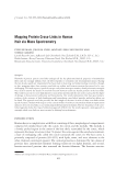
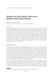
655 Address all correspondence to Evelyne Maes, evelyne.maes@agresearch.co.nz Mapping Protein Cross-Links in Human Hair via Mass Spectrometry EVELYNE MAES, JOLON M. DYER, SANTANU DEB-CHOUDHURY AND STEFAN CLERENS Lincoln Research Centre, AgResearch Ltd, Christchurch, New Zealand (E.M., J.M.D., S.D.-C., S.C.) Riddet Institute, Massey University, Palmerston North, New Zealand (E.M., J.M.D., S.C.) Biomolecular Interaction Centre, University of Canterbury, Christchurch, New Zealand (E.M., J.M.D., S.C.) Synopsis Networks of protein–protein cross-links underpin all the key physicomechanical properties of mammalian fibers and also strongly influence how the fiber responds to treatments and environmental insults. During the last decade, significant improvement has been made in detecting and understanding cross-links, though, to date, mapping where these protein cross-links are within and between proteins has proved to be very challenging. This work reports a proof-of-concept study where mass spectrometry–based proteomics strategies were used to unravel the details of cross-link location between trichocyte keratin proteins in the hair shaft. This work focuses on two cross-links known to exist in hair proteins and that are used as a proxy for the degree of damage in hair proteins: lanthionine and lysinoalanine. Our results demonstrate that mass spectrometric evidence of the existence of both lanthionine and lysinoalanine cross-linked peptides within hair fibers can be found. The approach used also provided the first insight of the exact location of these cross-links within specific keratins. Analysis with respect to the current models of trichocyte intermediate filament organization indicates detected cross-links occur within antiparallel keratin heterodimers. The low abundance of the cross- linked peptides makes this type of evaluation approach difficult, but the results represent a new approach to untangle which cross-links are most essential to defining hair performance. INTRODUCTION Human hair is a complex tissue with fibers consisting of three morphological compartments composed of cornified dead cells: the cuticle, the cortex, and the medulla. The medulla is a loosely packed region in the center of the hair shaft, surrounded by the cortex, which represents the major structure in hair. To protect the cortex against the outside environment, a layer of overlapping cells called the cuticle surrounds the cortex (1). The hair cortex is primarily composed of proteins (90–95%), with lipids, water, and trace elements as the other chemical constituents present. The cortical proteome is dominated by two classes of proteins: first, intermediate filament–forming trichocyte (or alpha) keratins, and second, keratin-associated proteins that surround the keratin filaments. The keratins associate J. Cosmet. Sci., 72, 655–669 (November/December 2021)

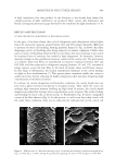
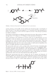
656 JOURNAL OF COSMETIC SCIENCE together to form coiled-coil dimers, intermediate filaments, and macrofibrils (2). To achieve a rigid hair structure, they form a strong compact network by both intra- and intermolecular interactions such as disulfide bonds, hydrogen bonds, electrostatic salt bonds, and amide bonds (3). Of major interest in hair science are the protein cross-links, such as disulfide bonds, which lead to the formation of covalent linkages between intermolecular amino acid residues and hence determine which regions within peptides and proteins are involved in the formation of protein networks. In addition to these native protein cross-links in hair, artificial cross-links, including lanthionine and lysinoalanine, can also be induced, mostly on insult such as heat, UV, or the use of alkaline products (4). Networks of protein–protein cross-links underpin all the key physicomechanical properties of mammalian fibers and strongly influence how the fiber responds to treatments and environmental insults. Understanding and controlling these networks is therefore critical to enhance our understanding of fiber science generally and develop new fiber treatments that enhance properties. However, due to the large degree of cross-linking present in mammalian fibers, analysis of these covalent bonds has been very difficult. Although some technologies have successfully led to the detection of cross-links (3), most techniques involve total protein destruction, making mapping where these cross-links are within and between proteins extremely difficult. In this proof-of-concept study, mass spectrometry–based proteomics strategies are used to unravel the details of cross-link location between trichocyte keratin proteins in the hair shaft. This work focuses on two cross-links known to exist in hair proteins and that are used as a proxy for the degree of damage in hair proteins: lanthionine and lysinoalanine. Both cross-links can be the result of degraded disulfide bonds to form dehydroalanine, or alternatively, of the elimination of water or hydrogen sulfide from serine and cysteine, respectively, to form dehydroalanine. This dehydroalanine intermediate will react with either lysine to form lysinoalanine or with cysteine to from lanthionine. Although mass spectrometry has been used before to elucidate protein cross-links in a food or medical context (5,6), this work, to our knowledge, is the first to decipher cross-links in the complex hair matrix with the aim of finding mass spectrometric evidence of the existence of cross-links within hair fibers and mapping their exact location within the protein in situ. METHODS SAMPLE PREPARATION Blended Caucasian virgin hair tresses were obtained from International Hair Importers & Products, Inc. (Glendale, NY, United States). Full-length fibers were cut into snippets of approximately 5 mm in length. For amino acid analysis, two types of samples were prepared: first, 10 mg of hair snippets were weighted, and second, hair protein extracts were prepared by using 100 mg of hair snippets, which were dissolved in 5 mL of an extraction buffer (7 M urea, 2 M thiourea, 0.05 M Tris, and 50 mM dithiothreitol (DTT), pH 7.5) at room temperature for 18 h using vigorous shaking. For proteomics analysis, 10 mg of hair snippets were dissolved in 1 mL of an extraction buffer (7 M urea, 2 M thiourea, 0.05 M Tris, and 50 mM DTT, pH 7.5) at room temperature for 18 h using vigorous shaking.
Purchased for the exclusive use of nofirst nolast (unknown)
From: SCC Media Library & Resource Center (library.scconline.org)