657 MAPPING PROTEIN CROSS-LINKS IN HUMAN HAIR PROTEIN DIGESTION After the protein extraction, an acetone precipitation was carried out to remove the extraction buffer components by adding 5 mL of ice-cold acetone to 1 mL of the sample. The samples were stored at –20°C for 48 h before they were centrifuged at 10,000 g and 4°C for 10 min. The protein pellet was washed twice with ice-cold acetone, air-dried, and resuspended in 200 μL of 10 mM ammonium bicarbonate. The protein concentration per sample was determined by Direct Detect (Millipore, MA, United States) according to manufacturer’s instructions. Next, 100 μg of each sample was aliquoted, and proteins were digested by sequencing grade trypsin (Promega) at a 1:50 ratio (trypsin:protein) for 18 h at 37°C at 300 rpm in a thermomixer. Next, the tryptic digest peptide mixtures were stored at –20°C until further analysis. AMINO ACID ANALYSIS The hair snippets and the tryptic digests were hydrolyzed with 6 N HCl at 110°C for 24 h. Amino acid analysis was performed using a Dionex HPLC system (Dionex UltiMate 3000 with Fluorescence Detector, Thermo Scientific, San Jose, CA, United States). Free amino acid residues and standards (containing lanthionine and lysinoalanine at a predetermined concentration) were derivatized with an AccQ-Tag reagent (Waters Corporation, Milford, MA, United States) and separated using a Thermo Accucore XL C18 column (4.6 mm i.d. × 250 mm, 4 μm particles) (Thermo Scientific, San Jose, CA, United States) at 37°C using a flow rate of 1.0 mL/min. Eluent A was AccQ-Tag solvent A, and eluent B was 100% acetonitrile. An excitation wavelength of 250 nm and emission wavelength of 395 nm were used for the quantitative analysis using a fluorescence detector (Dionex Ultimate FLD 3000, Thermo Scientific, San Jose, CA, United States). LIQUID CHROMATOGRAPHY–TANDEM MASS SPECTROMETRY To detect the cross-links via mass spectrometry, the peptide mixtures were separated on an Ultimate 3000 RSLCnano system (Thermo Scientific, San Jose, CA, United States) using a C18 PepMap100 nano-Trap column (200 μm i.d. × 2 cm) (Thermo Scientific, San Jose, CA, United States) connected to a ProntoSIL C18AQ analytical column (100 μm i.d. × 15 cm, 3 μm particle size, 200 A pore size) (nanoLCMS Solutions, Gold River, CA, United States). For each sample 1 μL (equal to 1 μg) was loaded on the trap column at 3 μL/min. The trap column was then switched in line with the analytical column. Reverse phase liquid chromatography (LC) separation was performed by a linear gradient of mobile phase B (0.1% formic acid in 100% acetonitrile) from 2% to 45% in 60 min, followed by a steep increase to 98% mobile phase B in 6 min, held at 95% mobile phase B for 2 min, returned to 2% mobile phase B over 5 min, and then re-equilibrated at 2% mobile phase B for 15 min resulting in a total run of 88 min used at a flow rate of 1,000 nL/min. The LC was coupled online to mass spectrometry via a Bruker CaptiveSpray ion source (Bruker Daltonics, Bremen, Germany) equipped with a nanobooster device and operated at 1,400 V. Data were acquired with a Q-TOF Impact II (Bruker Daltonics, Bremen, Germany) mass spectrometer in a dynamic data-dependent auto-MS/MS mode where a full scan spectrum (150–2,200 m/z, 2 Hz) was followed by a dynamic inclusion of collision-induced dissociation

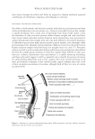
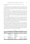
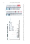
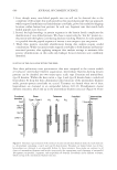
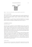
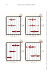
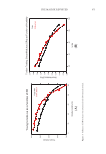
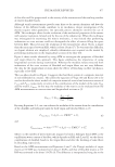
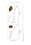
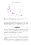
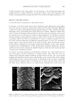
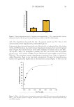
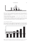
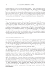
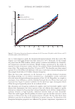
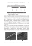
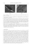
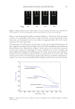
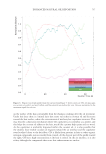
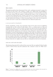
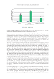
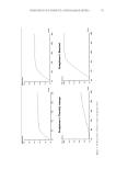
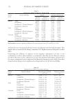
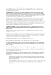
658 JOURNAL OF COSMETIC SCIENCE tandem mass spectra (2–32 Hz, depending on intensity) for 3 s. A preference of selection of multiple-charged precursors (2+ to 5+) was made, and an isolation width of 0.03 Th was set. Dynamic exclusion duration was set to 20 s. PROTEIN IDENTIFICATION Potential protein–protein cross-links were searched with Kojak v1.6 (7). The following parameters were used: a precursor mass tolerance of 10 ppm was allowed with fragment bin offset = 0, fragment bin size 0.03. A custom in-house database was created containing all human keratin protein sequences, keratin-associated proteins, and their reverse complements as decoy proteins. Trypsin was specified as a digestive enzyme, and up to three missed cleavages were allowed. A minimum peptide mass of 245 Da was set. Carbamidomethyl (C), oxidation (M), and deamidation (NQ) were chosen as variable modifications, and a maximum of three modifications per peptide was allowed. A lanthionine bond (Cys–Cys or Ser–Cys) or lysinoalanine bond (Lys–Cys) was specified as a cross-link. For each spectrum, the top 20 scoring peptides were used for cross- link combination. To minimize false positives, peptide spectrum matches exported from Kojak were further validated by Percolator V2.08 (8). The threshold for peptide spectrum matches after validation was set at q value smaller than 0.01. RESULTS CONFIRMATION OF CROSS-LINK PRESENCE IN HAIR FIBERS AND THEIR PROTEIN EXTRACTS Amino acid analysis was used to validate the presence of lysinoalanine or lanthionine cross-links in samples that were used for proteomics cross-link mapping. The amount of lanthionine and lysinoalanine present both in hair fiber snippets (one biological replicate run in duplicate) and hair snippet protein extracts (three separate protein extracts run in duplicate) is shown in Figure 1. Cross-links were detected in both the fiber and in the protein extract from the fiber, though some variability in the samples was noticed. Figure 1. The quantities of lanthionine and lysinoalanine cross-links (mg/100 mg) present in hair snippets (black) and triplicate hair extracts (blue) as determined by amino acid analysis represented with 95% confidence intervals.
Purchased for the exclusive use of nofirst nolast (unknown)
From: SCC Media Library & Resource Center (library.scconline.org)