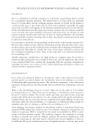
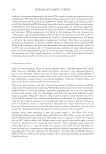
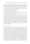
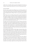
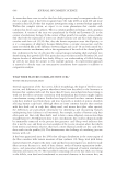
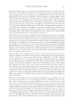
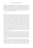
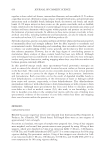
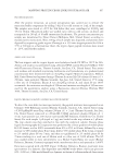
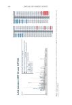
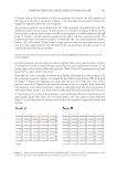
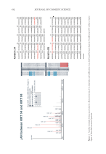
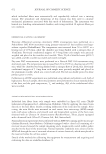
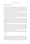
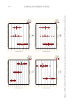
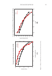
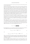
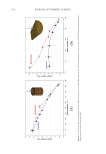
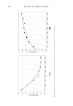
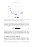
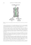
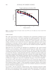
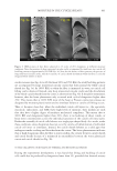
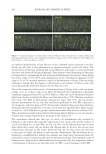
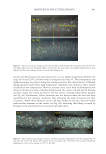
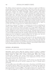
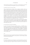
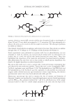
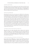
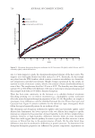
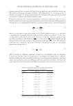
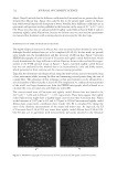
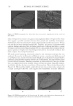
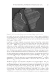
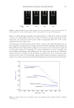
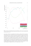
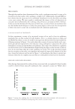
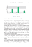
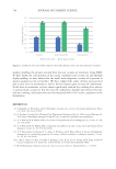
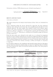
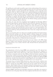
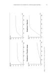
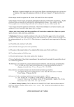
665 MAPPING PROTEIN CROSS-LINKS IN HUMAN HAIR unfold proteins to enable enzymatic digestion and mass spectrometric analysis (10). This proof-of-concept study focused on lanthionine and lysinoalanine cross-links induced by environmental wear and tear. To ensure that the target cross-links were present in our samples, amino acid analysis against lanthionine and lysinoalanine standards measured the absolute quantity of cross-links present in hairs and protein extracts. Although amino acid analysis did not indicate the molecular location of target cross-links, it was essential to creating a robust mass spectrometry and analysis method. In this work, mass spectrometric data acquisition and further analysis of the hair protein extracts did offer additional insights about the approach we developed for mapping cross-links: Figure 6. Protein sequences (human) of keratin 33B and keratin 80, with the peptide sequence involved in the cross-link indicated in red. Figure 7. Mass spectrometric characterization of intermolecule lysinoalanine cross-links found between keratin 34 and keratin 82 within a tryptic digest of hair shaft-extracted proteins. The signal-to-noise level of this cross-link is very low, and interpretation of the data should be done with caution as hardly any difference between the target ions and background ions is seen.

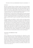
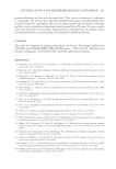
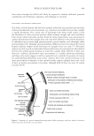
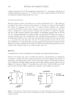
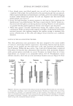
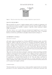
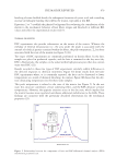
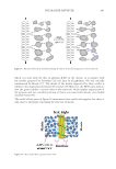
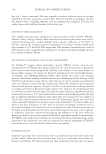
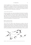
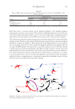
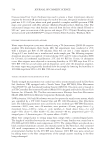
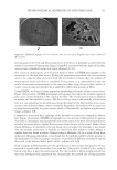
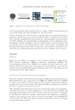
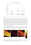
666 JOURNAL OF COSMETIC SCIENCE 1. First, though many cross-linked peptide ions can still not be detected due to the complexity of the sample, this study provides a first proof-of-principle that our approach, which targeted lanthionine and lysinoalanine cross-links, proved successful for mapping locations within human hair proteins. In each case, fragment ions that match both linked peptides were detected. 2. Second, the high homology in protein sequences in the keratin family complicates the identification of cross-linked proteins. We chose to report only the “first hit” protein (i.e., the protein with the highest score) during database matching. However, for some peptides it is possible that the peptide sequence of interest is not unique to just one protein. 3. Third, false positive cross-link identifications during data analysis require careful consideration. While our initial results mapped cross-links to both keratins and keratin- associated proteins, after applying stringent data analysis settings to minimize false positive identifications, in this work only linkages between keratins were confidently identified. POSITION OF THE CROSS-LINK WITHIN THE FIBER Next these preliminary mass spectrometric data were compared to the current models of trichocyte intermediate filament organization. Intermediate filament–forming keratin proteins can be classified into two major types: acidic type I keratins and neutral-basic type II keratins. Within the hair cortex, a type I and type II keratin form a coiled-coil heterodimer. To keep this three-dimensional coiled structure of the intermediate filament stable, protein–protein cross-links are crucial. Tetramers are formed when two of these heterodimers are clustered in an antiparallel fashion (9,11,12). Protofilaments cluster different tetramers, which end up in the intermediate filament structure (Figure 8). Three Figure 8. Schematic representation of the trichocyte keratin intermediate filament molecule. Each molecule is a heterodimer containing a type I and type II chain keratin. Tetramers are formed when two of these heterodimers are clustered in an antiparallel fashion. In these tetramers, three modes are common to all classes of intermediate filaments and involve (respectively) the approximate axial alignment of antiparallel 1B segments, antiparallel 2 segments, and antiparallel rod domains. Protofibrils cluster different tetramers, which end up in the intermediate filament structure.
Purchased for the exclusive use of nofirst nolast (unknown)
From: SCC Media Library & Resource Center (library.scconline.org)