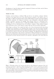
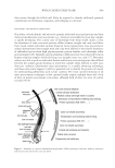
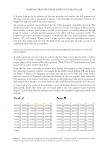
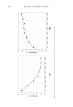
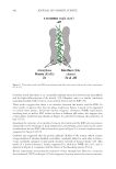
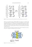
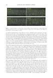
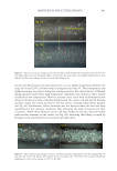
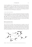
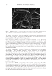
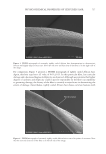
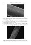
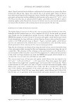
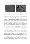
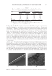
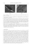
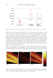
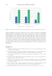
713 PHYSICOCHEMICAL PROPERTIES OF TEXTURED HAIR MACROPHOTOGRAPHY OF HAIR Photographic images of hair were obtained using an Epson Perfection V550 Photo Color Scanner (Epson Seiko, Suwa, Japan) with Epson Scan (version 3.9.1.1US) software. Images were collected in 24-bit color mode at 300 dpi. Image analysis of the photographs was carried out with ImageJ (version 1.53a) software (National Institutes of Health, Bethesda, MD). The lengths of individual hair fibers in the relaxed and fully extended states were measured to determine the curl index (CI) of hair. HAIR CROSS-SECTION PREPARATION Hair obtained from tresses was sectioned using a Leica CM3050 S (Leica Microsystems GmbH, Wetzlar, Germany) cryostat equipped with a high-profile sectioning head and Leica 818 high-profile cutting blades. Note that the cryostat blade was changed after each sample. For each sample, a 25 mm aluminum specimen disk was pre-equilibrated in dry ice (−78.5°C). A small section (1/8–1/4 in. wide × 1–2 in. long) of the ¾ in. wide tress was removed from the center of the tress length. The tip-end of the damp fiber section was then held straight and perpendicular against the platform of the disk. At the interface between the disk and tip-end of the fiber bundle, a drop of distilled water was then added. The drop instantly crystallized, hence causing the tip-end of the fiber bundle to adhere to the aluminum specimen disk. By pulling gently upward on the root end of the fibers and slowly adding water to the fiber bundle, a straight rod of ice-embedded hair was produced. Separately, each embedded sample was then mounted onto the specimen head (−30°C) of the cryostat, where the chamber temperature was equilibrated at −25°C. After conditioning for at least 1 h, 5-μm–thick sections were collected in continuous mode using a sectioning speed of 60% maximum. Hair cross-sections were air-dried on paper overnight under reduced pressure. FIELD EMISSION SCANNING ELECTRON MICROSCOPY (FESEM) OF HAIR The cross-sections were imaged using field emission scanning electron microscopy (FESEM) to investigate the internal morphological structure of hair and to calculate the cortical cross-sectional area (n=100). Cross-sections were fixed to aluminum Pelco pin stubs using 25 mm conductive carbon tabs (Ted Pella, Redding, CA) and then coated with Au/Pd using our Leica EM ACE600 sputter coater. Finally, the hair fiber cross-sections were imaged with a Hitachi SU-5000 FESEM (Hitachi High Technologies, Schaumburg, IL) using various magnifications. FOURIER TRANSFORM INFRARED (FTIR) IMAGING OF HAIR CROSS-SECTIONS Fourier transform infrared (FTIR) images were obtained with a PerkinElmer Spotlight 400 FTIR imaging microscope (PerkinElmer Inc., Waltham, MA), which combines an optical microscope with an FTIR spectrometer. The system consists of a linear array of mercury- cadmium-telluride detectors coupled to a precision automated X–Y sampling stage. Background spectra were collected on sample-free areas of the CaF 2 crystal, and FTIR images were obtained at 8 cm−1 spectral resolution in transmittance mode at 16 scans/pixel.

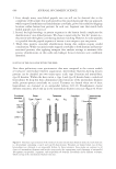
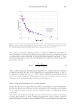
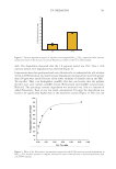
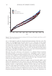
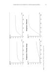
714 JOURNAL OF COSMETIC SCIENCE Cross-sectioned hair (5 μm thickness) was used to ensure a linear transmission detector response for the entire IR spectral range. For each of the scans, the spatial resolution of each pixel was 6.25 × 6.25 μm, where each pixel provided a complete mid-IR spectrum. FTIR maps were generated with ISys software (Malvern Panalytical Ltd., Malvern, UK). The concatenated images were then baseline corrected from the base of the Amide II band to 900 cm−1 prior to truncation of the spectra and images (150 × 150 μm). Resulting spectra were processed with GRAMS/AI™ software (Thermo Fisher Scientific, Waltham, MA). DYNAMIC VAPOR SORPTION (DVS) OF HAIR Water vapor absorption curves were obtained using a TA Instruments Q5000 SA sorption analyzer (TA Instruments, New Castle, DE). All experiments were conducted at 25˚C with a nitrogen gas flow of 200 mL/min. Hair was cut into 1–2 mm snippets, and 10 mg ± 0.5 was loaded into a stainless-steel mesh sample pan. The following sorption- desorption procedure was applied: (1) initial drying: 60˚C and 0% relative humidity (RH) for 120 min (2) isothermal equilibration: 25˚C and 0% RH for 15 min (3) absorption curve: fiber snippets were subjected to increasing humidity in 10% RH steps from 0% to 90% RH (720 min at each step) and (4) desorption curve: after the absorption sequence, the water vapor was progressively desorbed from the sample by lowering the humidity in 10% RH steps from 90% to 0% RH (720 min at each step). TENSILE STRENGTH MEASUREMENTS OF HAIR Tensile strength measurements were carried out with a system manufactured by Dia-Stron Ltd. (Andover, UK) equipped with a Tensile Tester (Type MTT690), Fiber Micrometer (Type FDAS770), and Automated Loading System (ALS1500). The entire unit is housed in an ETS Controlled Environment Chamber (Model 5533) designed and built by Electro-Tech Systems, Inc. (Glenside, PA). The fiber micrometer was a Mitutoyo Laser Scan Micrometer (Model LSM-6200) from Mitutoyo Corporation (Kawasaki, Japan). UvWin (version 3.60, build 8) software was used to operate the tensile tester system, which was controlled by a UV 1000 Control Unit and PU 1100 Pneumatics Unit (Dia-Stron Ltd.). The following parameters were used for the tests: method type–MTT680 Extension extension—100%, mm rate—20 mm/min start position—30 mm gauge force—3 g maximum force—2,000 g cycles—1 cycle number—1 break detect force—5 g end angle—360 slices—1 filter width—32 data interval—80 ms proportional gain—25 integral gain—2 derivative gain—50 system offset—24,650. Fibers were crimped prior to testing using brass crimps with a custom-designed press. The length of each fiber between the two crimps was 30 mm. Studies were carried out at 65% RH and 100% RH at room temperature (22°C). The crimped fibers were placed on a black anodized aluminum carousel, also manufactured by Dia-Stron Ltd., that contained 100 small compartments to house individual fibers. Humidity was controlled by the environmental chamber for the 65% RH tests however, 100% RH tests were carried out by adding water to the fiber compartments of the sample cassette resulting in the fiber being submerged.
Purchased for the exclusive use of nofirst nolast (unknown)
From: SCC Media Library & Resource Center (library.scconline.org)