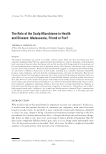
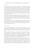
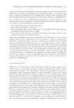
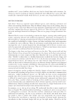
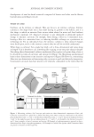
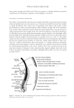
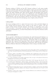
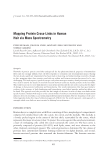
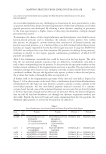
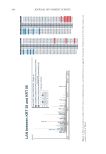
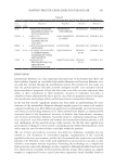
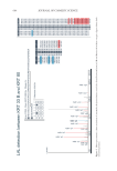
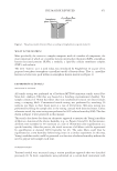
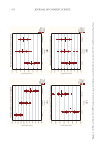
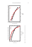
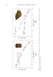
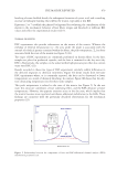
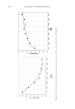
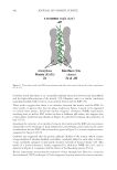
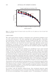
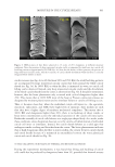
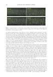
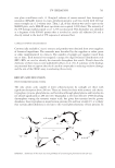
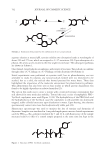
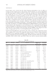
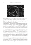
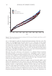
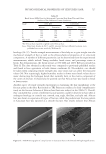
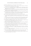
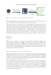
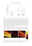
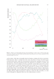
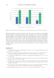
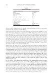
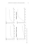
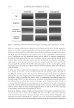
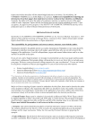
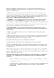
661 MAPPING PROTEIN CROSS-LINKS IN HUMAN HAIR A further look at the localization of the two peptides (red) within the full sequences of the respective proteins is visualized in Figure 3. Interestingly, the peptides of interest are mapped at opposite ends of the total sequence. An analogous analysis was performed for the other potential cross-links detected. The lanthionine link between keratin 34 and keratin 85 is visualized in Figure 4. Similar to the first example, no direct proof of the cross-link via a fragment ion could be detected, though proof of existence of both peptide sequences (the alpha and beta sequence) within the fragment spectrum can be found (Figure 4). Although the cross-linked peptides between keratin 33A and keratin 74 were found in two spectra, most ion intensities were again very low this might indicate that the bulk of the ion intensities are still too low to be confidently detectable by our method. DETECTION OF LYSINOALANINE CROSS-LINKS WITHIN HAIR FIBER PROTEIN EXTRACTS VIA MASS SPECTROMETRY A similar approach was also taken to explore whether there is mass spectrometric evidence of lysinoalanine bonds between keratins and/or keratin-associated proteins present in the tryptic digest of the extracted hair fiber proteins. Table II lists all lysinoalanine cross-links found with q values smaller than 0.05. From this list, two cross-links of interest were further investigated on their potential. At first, mass spectrometric evidence was sought for the link between keratin 33B and keratin 80. Figure 5 displays the fragment spectrum with the detected b-ions and y-ions in blue and red, respectively. Figure 6 represents the location of the two peptides (red) within the full sequences of the respective proteins. Similar to the lanthionine data, proof of presence of both peptides was found as ions from both the alpha peptide and beta peptide were detected. A closer look at the potential lysinoalanine cross-link between keratin 34 and keratin 82, alternatively, shows that most ions are found with a very low signal-to-noise threshold (Figure 7), indicating that the interpretation of these data should be approached with extra caution. Figure 3. Protein sequences (human) of keratin 38 and keratin 85, with the peptide sequence involved in the cross-link indicated in red, the head domain underlined, and the tail domain in italics.

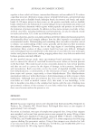
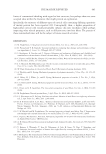
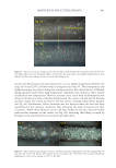
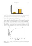
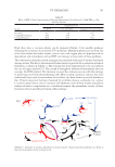
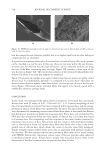
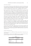
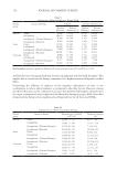
Figure 4. Another example of mass spectrometric characterization of an intermolecule lanthionine cross-link found between keratin 34 and keratin 85 within a tryptic digest of hair shaft-extracted proteins. 662 JOURNAL OF COSMETIC SCIENCE
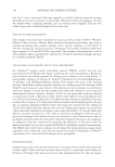
Purchased for the exclusive use of nofirst nolast (unknown)
From: SCC Media Library & Resource Center (library.scconline.org)