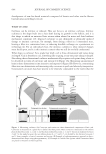
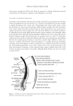
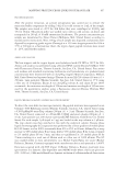
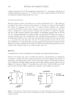
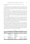
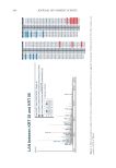
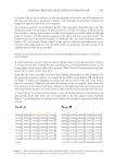
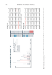
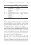
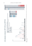
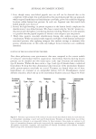
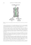
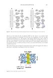
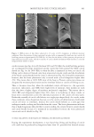
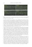
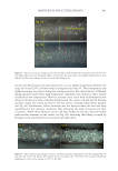
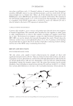
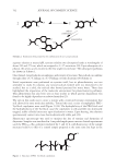
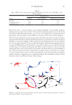
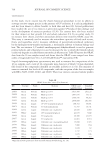
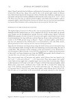
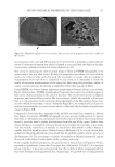
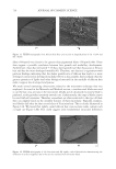
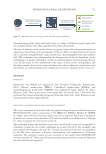
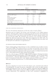
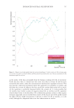
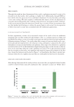
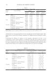
667 MAPPING PROTEIN CROSS-LINKS IN HUMAN HAIR different modes of molecular assembly of intermediate filaments are described. These three modes are common to all classes of intermediate filaments and involve (respectively) the approximate axial alignment of antiparallel 1B segments (model A 11 ), antiparallel 2 segments (model A 22 ), and antiparallel rod domains (model A 12 ) (Figure 8). Previous work from the Parry et al. research groups (11,13–16) do show that newly assembled protofilament molecules associate in A 11 or A 22 alignments within the follicle before a profound structural rearrangement occurs when the environment changes from a reducing to an oxidizing one. We compared these filament models with our experimental data to get a better idea of what these positions of the cross-linked peptides within the protein sequence mean, and to see if our data aligned with the models of trichocyte intermediate filament organization. Important to note, though, is that this work is not intending to make any assumptions on the structural intermediate filament biology aspect. The lanthionine cross-link found between keratin 38 and keratin 85 matches the models in a number of ways: first, proteins keratin 38 and keratin 85 are, respectively, a type I and type II keratin and could thus form a heterodimer. They are both cortex proteins. Second, the position of the cross-linked peptides (Figure 3) demonstrates that the link is made between proteins in antiparallel positioned dimers forming a tetramer, with an interaction between a head domain of one protein and a tail domain of another. This could be an indication of the suggested model A 12 in a tetramer but could also be the packaging of adjacent tetramers. Third, the A 12 arrangement is probably the most prevalent in a mature hair fiber as it lends itself to optimal molecular packing and radial compaction in an oxidized environment (13). Subsequently, we researched whether the other detected lanthionine cross-links could also be explained by these models. Similar to the previous example, the cross-link between keratin 33A and keratin 74, detected in two spectra (Table I), did show an A 12 arrangement, with peptide 1 (AA 91–108) from keratin 33A located in linker 1, while peptide 2 from keratin 74 with amino acid position 370–378 in the sequence is part of coil 2. As these two proteins do not tend to be found in the same part of the fiber (i.e., K33a is in the cortex and medulla, while K74 is found in the inner root sheet), it is still a question of where this cross-link would be formed, as both peptides are unique and thus only identified in these respective proteins. The mass spectrometric evidence of the lanthionine cross-link between keratin 34 and keratin 85, a type I and type II keratin, does show a different positioning of the cross-linked peptides. Here, one peptide is found at the N-terminus (head region), while the other peptide is found in the linker 1 region (Figure 5). This lanthionine link would thus suggest that these two proteins are more comparable to the A 11 model, though the position of the head sequence in the intermediate filament is still questionable. Similarities between the proposed intermediate filament models are also found when looking at the trichocyte keratin lysinoalanine cross-links within the samples. Mass spectrometric mapping of a lysinoalanine cross-link between keratin 33B and keratin 80 displays an interesting link, as both proteins are present in the medulla. The medulla, however, is known to have a heterogenous structure, and hence, although the position of the peptides of interest shows that these are somewhat comparable to model A 22 , where the proteins are still present in antiparallel heterodimers within the filament, these may or may not form tetramers in the medulla. Though some mass spectrometric evidence for the presence of the other detected lysinoalanine cross-links is found, manual inspection of the data (e.g., the cross-link between keratin 34

668 JOURNAL OF COSMETIC SCIENCE and keratin 82 [Figure 7]) shows that the peptides are detected with fragments of very low intensity, and a distinction between the fragment ions and the background signals is hard to detect. Therefore, none of the other lysinoalanine data were taken forward for further comparison against the intermediate filament models. In conclusion, our results demonstrate here, for the first time, the mass spectrometric characterization of native cross-links within hair fibers (lanthionine and lysinoalanine), combined with their mapping within the protein sequence, and their potential mapping within the intermediate filament. These results represent the first steps toward untangling the many unknowns related to cross-links in fibers. However, caution should still be taken, as our results clearly indicate that the low abundance of the cross-linked peptides still makes this type of evaluation approach difficult, even when optimized mass spectrometric data acquisition methods are applied. These preliminary data, however, do show evidence of previous reported heterodimer compositions and do showcase the potential of the technology. ACKNOWLEDGMENTS We would like to thank Dr. Duane Harland and Dr. Jeffrey Plowman for their critical input in this work. This work was partially funded and supported by a contract with the Procter & Gamble Company and by the AgResearch Strategic Science Investment Fund. REFERENCES (1) C. R. Robbins, “Morphological, macromolecular structure and hair growth,” Chemical and Physical Behavior of Human Hair (Springer, Berlin, Heidelberg, 2012), pp. 1–104. (2) D. P. Harland, R. J. Walls, J. A. Vernon, J. M. Dyer, J. L. Woods, and F. Bell, Three-dimensional architecture of macrofibrils in the human scalp hair cortex, J. Struct. Biol., 185(3), 397–404 (2014). (3) S. Deb-Choudhury, Crosslinking between trichocyte keratins and keratin-associated proteins, Adv. Exp. Med. Biol., 1054, 173–183 (2018). (4) A. L. Miranda-Vilela, A. J. Botelho, and L. A. Muehlmann, An overview of chemical straightening of human hair: technical aspects, potential risks to hair fibre and health and legal issues, Int. J. Cosmet. Sci., 36(1), 2–11 (2014). (5) E. Maes, J. M. Dyer, H. J. McKerchar, S. Deb-Choudhury, and S. Clerens, Protein–protein cross-linking and human health: the challenge of elucidating with mass spectrometry, Expert Rev. Proteomics, 14(10), 917–929 (2017). (6) H. J. McKerchar, S. Clerens, R. C. J. Dobson, J. M. Dyer, E. Maes, and J. A. Gerrard, Protein–protein crosslinking in food: proteomic characterisation methods, consequences and applications, Trends in Food Science & Technology, 86, 217–229 (2019). (7) M. R. Hoopmann, A. Zelter, R. S. Johnson, M. Riffle, M. J. MacCoss, T. N. Davis, and R. L. Moritz, Kojak: efficient analysis of chemically cross-linked protein complexes, J. Proteome Res., 14(5), 2190–2198 (2015). (8) L. Kall, J. D. Canterbury, J. Weston, W. S. Noble, and M. J. MacCoss, Semi-supervised learning for peptide identification from shotgun proteomics datasets, Nat. Methods, 4(11), 923–925 (2007). (9) R. D. B. Fraser and D. A. D. Parry, Structural hierarchy of trichocyte keratin intermediate filaments, Adv. Exp. Med. Biol., 1054, 57–70 (2018). (10) E. Maes, S. Clerens, J. M. Dyer, and S. Deb-Choudhury, Improved detection and fragmentation of disulphide-linked peptides, Methods Protoc., 1(3), (2018).
Purchased for the exclusive use of nofirst nolast (unknown)
From: SCC Media Library & Resource Center (library.scconline.org)