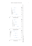
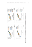
33 Silver Nanoparticles of Bee Honey the refractive index at 20°C. The pH value was determined by a pH meter on a solution composed of 10 g of bee honey and 75 ml of distilled water. Electrical conductivity was measured (in μS/cm) using a conductometer device at 20°C of the test solution that consisted of 20% bee honey weighed as dry matter dissolved in distilled water and brought to a volume of 1/5. BIOSYNTHESIS OF AgNPs In the silver nanoparticles biosynthesis, 100 ml of freshly prepared solution of 1 mM AgNO 3 was added to 100 g bee honey in 250-ml Erlenmeyer flasks. The composition solution was stirred and incubated for 8 hours. After incubation, the mixture containing the nanoparticles was centrifuged and the pellets were lyophilized. The lyophilized AgNPs–bee honey nanoparticles were stored for further analysis. The samples of AgNPs– bee honey nanoparticles (A, B, and C) were bee honey from longan flowers, wildflowers, and benjaphan flowers, respectively. CHARACTERIZATION OF AGNPS–BEE HONEY NANOPARTICLES UV-visible spectroscopy. The nanoparticles of AgNPs–bee honey nanoparticles were demonstrated using a UV-visible spectrophotometer by taking absorption samples at 200 to 800 nm after 30 minutes to 8 hours incubation. FTIR analysis. FTIR analysis of AgNPs–bee honey nanoparticles was performed using a durable diamond single-reflection mode. The spectrum was recorded using Thermo Scientific (Nicolet IS5) FT-IR spectroscopy using transmittance mode and operating with 4 cm−1 correction. Particle size distribution. Particle size distribution is measured from a tangible material provided by nanoparticles. The size distribution profile of AgNPs–bee honey nanoparticles was measured using dynamic light scattering (DLS) (Malvern Panalytical Ltd., Malvern, UK). Water samples of the nanoparticles (5 ml) were diluted with double distilled water (50 ml) using sodium chloride as electrolyte suspending solution (2 × 10⁻² M NaCl). The pH was then adjusted to the required value. Samples were stirred for 30 minutes. After stirring, the measuring pH was recorded, and the particle size distribution of the metal particles was measured. In each case, an average of three ratings was reported. ANTIOXIDATION ACTIVITY DPPH–free radical scavenging. The antiradical activity of bee honey samples was measured according to the procedure of Ali et al. (9). A bee honey sample was dissolved in distilled water in concentrations from 125 to 500 mg/ml, and 0.2 ml of each solution was combined with 1.8 ml of 130 µM DPPH (final concentration 83.3 µM) dissolved in ethanol complete with 1 ml of acetate buffer solution (100 mM, pH 5.5). The mixtures were stirred vigorously and left for 30 minutes at room temperature in the dark, after which the remaining DPPH absorption was measured at 517 nm compared to blank to eliminate the effect of bee honey color. The blank sample was bee honey, in the same concentration as described above, and acetate buffer without DPPH solution. In each bee honey concentration tested, the

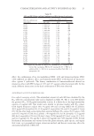
34 JOURNAL OF COSMETIC SCIENCE remaining DPPH percentage was calculated. The percent scavenging activity of DPPH was calculated using the following formula: %Scavenging [(1- BS/BC)]×100 =where “BS” and “BC” correspond to absorbance of sample and absorbance of control, respectively. The result was expressed in equivalent μg butylated hydroxytoluene per 1 g of dried sample. Ferric reducing/antioxidant power assay (FRAP). The procedure described by Benzie et al. was used (10). The goal of this approach is based on the reduction of a ferric 2,4,6-tripyridyl- s-triazine complex (Fe3+-TPTZ) to its ferrous, colored form (Fe2+-TPTZ) in the presence of antioxidants. The FRAP reagent contained 2.5 ml of a 10 mM TPTZ (2,4,6-tripyridyl- s-triazine) solution in 40 mM HCl, 2.5 ml of 20 mM FeCl 3 ,and 25 ml of 0.3 M acetate buffer, pH 3.6. It was prepared daily and warmed to 37°C. Aliquots of 200 µL of sample were mixed with 1.8 ml of FRAP reagent, and the absorbance of the reaction mixture was measured at 593 nm by spectrophotometer after incorporated at 37°C for 10 minutes compared to the sugar analog. The aqueous standard solutions of FeSO 4 .7H 2 O (100– 1,000 µM) were used for the calibration curve, and the results were expressed as the FRAP value (µM Fe(II)) of the bee honey sample solution. ANTIMICROBIAL ACTIVITY The antimicrobial susceptibility of AgNPs–bee honey and bee honey were evaluated using the disk diffusion method. The stability of disks containing oxacillin and gentamicin was prepared. Disposable plates were incubated with the tested gram-positive (S aureus) and gram-negative (Escherichia coli) bacteria. The antibacterial effect of AgNPs–bee honey was tested and evaluated against bacteria where 2.0 × 10⁸ CFU/ml by determining the minimum inhibitory detection (11) on microdilution plates using Mueller Hinton Broth (Difco™). The concentrations of AgNPs–bee honey and bee honey (AgNO 3 used as a control) ranged from 15.62 to 1,000 µg/ml. Different concentrations (15.62, 31.25, 62.50, 125.00, 250.00, 500.00, and 1,000 µg/ml) of AgNPs were dispensed into each well. Zones of inhibition were measured after 24 hours of incubation at 37°C. SERUM FORMULATION To prepare 100 g serum, 2.0 g of glycerin, 0.25 g of tween 80, and 0.25 g of poly (ethylene glycol) were added and mixed in a water bath at 80°C for the oil phase. Then 0.5 g of xanthan gum and 0.1 g methyl paraben were dissolved in 94.9 ml of distilled water at 50°C to prepare the liquid phase. The liquid phase was used to dissolve 2.0 g of AgNPs– bee honey nanoparticles, and the mixture was added slowly with continuous stirring in the oil phase to form a cold silver nanoparticles serum. Another cold cream without the nanoparticles was also formulated to serve as a control (Table I).
Purchased for the exclusive use of nofirst nolast (unknown)
From: SCC Media Library & Resource Center (library.scconline.org)